November/December 2024 competitors

“Seeing is believing.” This freely suspended structure was fabricated by two-photon lithography (TPL), followed by atomic layer deposition (ALD) of Ni on a SiN membrane frame without a membrane. The free-form design achieved by TPL and the 3D conformal coating by ALD are being seen. Microscope: SEM Gemini.

InP grown by MBE turns contamination into fascinating art – shaping intricate, fortress-like layers that echo medieval defenses. Microscope: SEM Merlin.

The packing of graphene layers forms a flower carpet. The picture was taken in TEM-Spectra.

This SEM image of crystalline, patterned depositions of silica on graphite, resembles blooming floral clusters. Microscope: SEM Teneo.

This is part of a contamination particle found on my sample. It could be organic or just a piece of the carbon tape. Microscope: SEM Gemini.

The polished cross-section of a GaN wafer. With the “Depth of Field” column mode on the SEM Merlin, one can observe the roughness of the wafer’s backside, coming from the original cutting with a wire saw. It reminds of a panorama of the Alps. Microscope: SEM Merlin.

A lake diatom collected in front of Epfl. Diatoms are single cell photosynthetic algae. Their protective frustules (shell) consists of porous siliceous material. Micro and nano pores will optimize light harvesting and allow exchange of nutrients . Diatoms play a major role in ocean’s oxygene production and in fixing carbon. They attract interest for their photonic properties or many applications in filtering , drug delivery, biosensors or bio-inspired materials. Gold-palladium coated, shot at 3 kV. Microscope: SEM Gemini.

The carbon membrane on Cu cracks in a way similar to running rivers. Microscope: SEM Teneo.

It should be a metal scrap attached to a gold contact during the lift-off process. Its brightness changed while observing it. Microscope: SEM Gemini.

This SEM image of the broken silica film on graphite resembles a network of dried riverbeds or cracked earth, evoking a natural, fractured landscape on a microscopic scale. Microscope: SEM Teneo.

This pattern of fractured silica film on HOPG resembles a patchwork of dried riverbeds. Microscope: SEM Teneo.

This stunning SEM image captures a strikingly bear-like structure perched atop a forest of porous silicon nanostructures, with other faint figures lurking in the background. Microscope: SEM Gemini.

This stunning SEM image captures a strikingly bear-like structure perched atop a forest of porous silicon nanostructures, with other faint figures lurking in the background. Microscope: SEM Gemini.

This stunning SEM image captures a strikingly bear-like structure perched atop a forest of porous silicon nanostructures, with other faint figures lurking in the background. Microscope: SEM Gemini.

Photographing the ancestor of microscopes through the lens of a modern instrument A cluster of Gallium nanoparticles resembling a magnifier. The layering of particles and the presence of subtle gallium oxide impurities give rise to a 3D effect reminiscent of a crafted lens. Captured by Spectra200 S/TEM in EPFL Valais

The cross section of fractured polyethylenimine fibres look like huge waves blown by strong winds. Microscope: SEM Gemini.

Ruptured thermoplastic microspheres left characteristic imprints in another thermoplastic matrix. Microscope: SEM Merlin.
September/October 2024 competitors

The glimpse of a small part of cross section from a PC polymer micro fibre looks like the Goddess of Victory statue displayed in the Louvre Museum. Microscope: SEM Gemini.

Ni foam support used in water splitting that looks like a sad, hanging ghost. Microscope: SEM Gemini.

Cu-Ag core-shell particles Microscope: TEM Osiris.

COF particles were deposited on Si wafer. The COF particles clustered into ‘rock-like’ formations, while the flat surface of the Si wafer resembled a ‘river’. Microscope: SEM Teneo.

Arsenic triselenide droplet that has stuck to the wafer side and is charging a lot, looking like a pancake being flipped, still steaming. Microscope: SEM Gemini.
July/August 2024 competitors

Broken arsenic selenide nanowire array caused by elastic wave looks like an earthquake. Microscope: SEM Gemini.

Salt dendrites with polymer nanoparticles. Observed by TEM. Microscope: TEM Talos.

Living in Switzerland means loving the outdoor as hiking. Now imagine hiking up a mountain while seeing thunderclouds above it. Scary, isnt’t it? Luckily in the microscale it is much less frightening. This micro-Matterhorn is a piece of Silicon with a glass backside (which accumulated the charges looking like a thunderstorm). Microscope: SEM Gemini.

SEM side view of a dewetted arsenic triselenide thin film on a flat substrate, showing typical uniform microscale droplets as well as smaller droplets in chemically modified regions. Microscope: SEM Gemini.

Nanograss—a cupric oxide (CuO) layer grown on a Si wafer via oxidizing a thin Cu layer. Microscope: SEM Merlin.

Nanograss—a cupric oxide (CuO) layer grown on a Si wafer via oxidizing a thin Cu layer. Microscope: SEM Merlin.
May/June 2024 competitors

After a rainy spring, sunlight allows flowers to grow back, on an Al microelectrode. Microscope: TEM Bio – Talos.

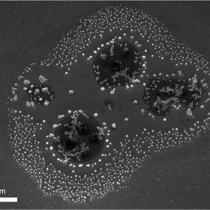
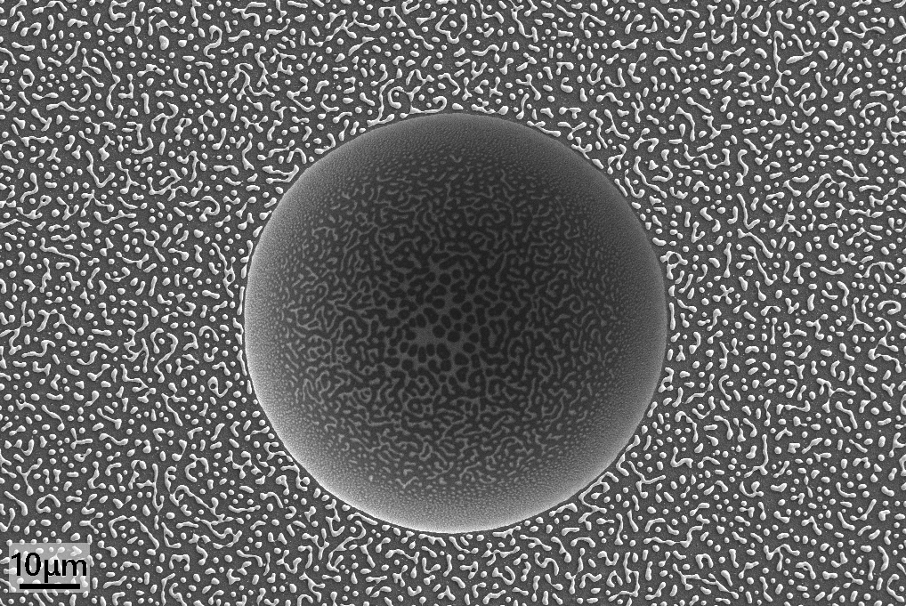
Dewetted arsenic triselenide thin film on a flat substrate, showing typical uniform microscale droplets as well as smaller droplets in chemically modified regions. Microscope: SEM Gemini.

The image looks quite like a goldfish with a human face. Microscope: SEM Teneo.

Curved polyethylenimine nanosheets look like broken ankle shackles on a foot(you can even distinguish toes), symbolizing the pursuit of freedom. Microscope: SEM Gemini.

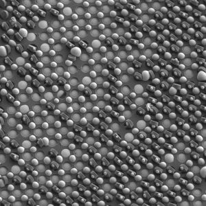
Carbon nanotubes on the electrode. Microscope: SEM Gemini.

3…2…1… Smile! Your picture is being taken 🙂 The alumina capping on my GeSn microstructures cracked into this camera-lens shape during annealing. Microscope: SEM Merlin.

SEM side view of a dewetted arsenic triselenide thin film on a flat substrate, showing typical uniform microscale droplets as well as smaller droplets in chemically modified regions. Microscope: SEM Gemini.

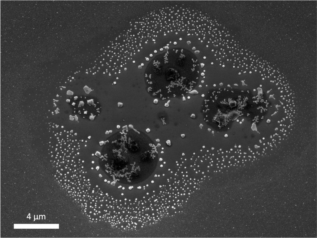
It was a sample of SrTiO3 epitaxial membrane stamped on a Nb doped SrTiO3 substrate. The chunk was prepared using Nvision then the whole sample underwent a thermal treatment under atmospheric pressure. The thermal treatment has completely recrystallised the sample with the implemented Ga to form different forms of oxides. These new grains formed an image of doves in the sample after thinning down. Microscope: SEM/FIB NVision.
March/April 2024 competitors

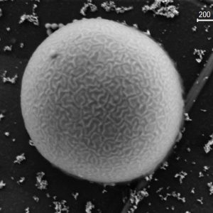
SEM image of small As2Se3 structures that slipped out from their mould, looking like small cupcakes with some topping on them. Microscope: SEM Gemini.

Kelp-like structures inside hydrogels. Microscope: SEM Gemini.

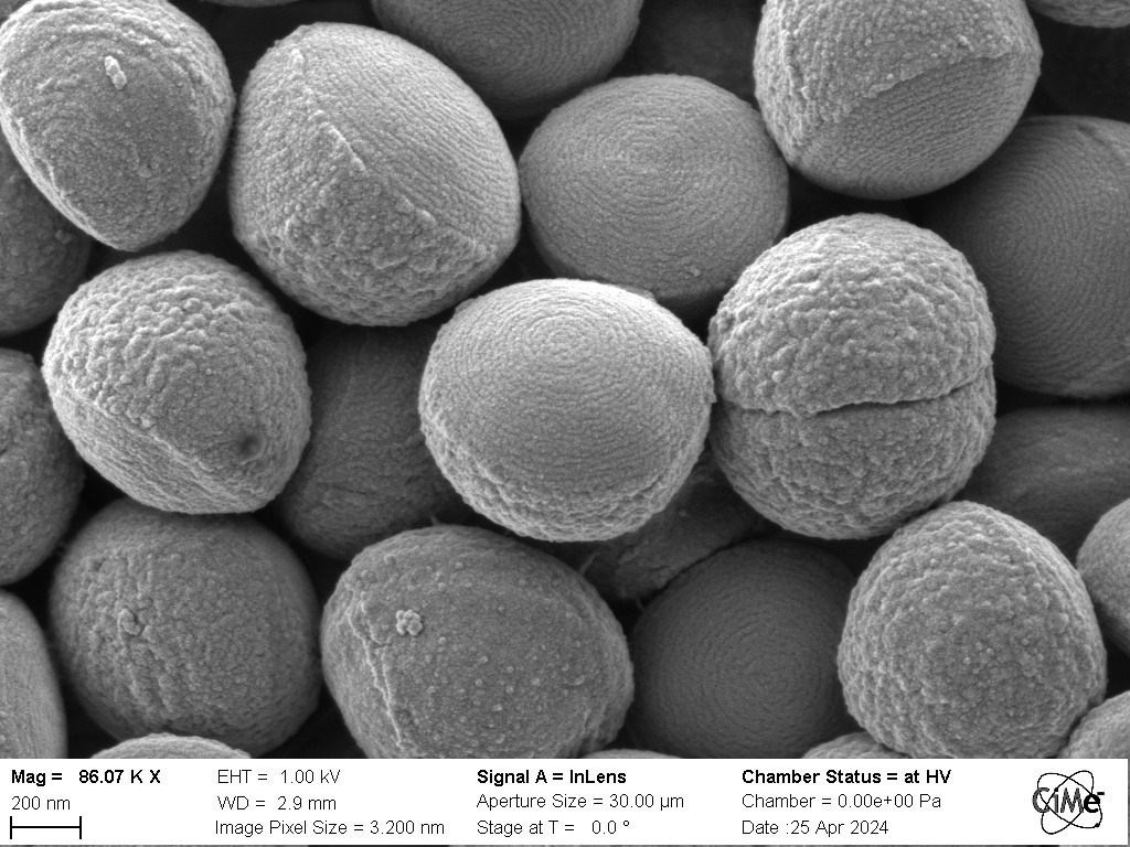
SEM image of biscuits made of Staphylococcus Xylosus. Hopefully we don’t eat too many of them… Sample greatly prepared by Mary-Claude Croisier-Coeytaux. Microscope: SEM Gemini.

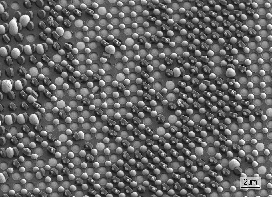
Bowl-shape polymer particles surrounded by other micro and polymer nanoparticles. Microscope: SEM Gemini.

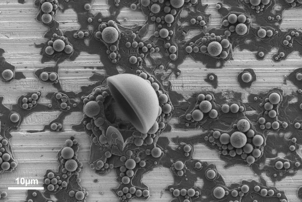
A bad supply chain management caused problems during the unmoulding and decoration of As2Se3 cupcakes, resulting in a huge chaos at the bakery. Microscope: SEM Gemini.

Gas Diffusion Electrode after electrochemical CO2 reduction. Microscope: SEM Teneo.

Covalent Organic Frameworks (COFs) look like a nanoscale coral forest growing on the surface of a cellulose nanofiber (CNF) film when viewed from above. Microscope: SEM Merlin.

Wild-type Synechocystis sp. PCC6803 cyanobacterium immobilized and fixed on a graphite foil support. Microscope: SEM Merlin.

Conductive polymer (PEDOT) growing on carbon fibers in a mesh (carbon felt). Microscope: SEM Gemini.
January/February 2024 competitors

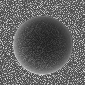
Dreaming of a tropical holiday in February? Me too. Join me for an exclusive excursion to these (contamination) nanoislands in a sea of germanium tin! Price: interest in the science. Microscope: SEM Merlin.

An undesirable air bubble created a hole in a flat substrate, which turned into a dark moon hiding the sun in a starry sky after the dewetting of a thin selenium film. Microscope: SEM Gemini.

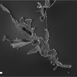
What do you see in this picture? Gecko? Warrior? A person walking their dog? Everyone has seen something different in this little metallic flake… what does this say about you? Microscope: SEM Merlin.

February is the month of love, and for you, CIME, I want to give a µ-rose. This is a beautiful mistake that resulted from contamination on my germanium tin film. Thank you to CIME for all that you do! Microscope: SEM Merlin.

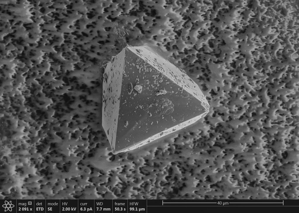
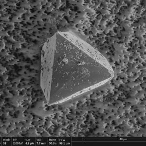
A single crystal of UiO-68 MOFs analogue, looks like an abandoned pyramid covered in a thick layer of sand. Microscope: SEM Teneo.

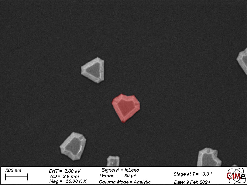
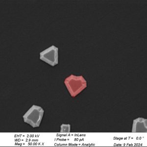
Zinc phosphite grown on graphene by MBE tends to grow as triangles. By chance two grains merged to form the perfect little heart. Microscope: SEM Merlin.

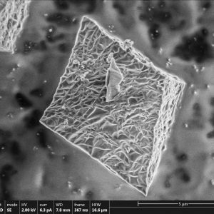
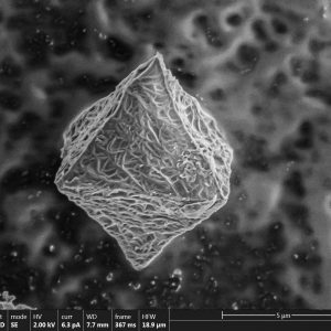
A single crystal of UiO-68 MOFs analogue with a rough surface, which makes it looks like cocoon or bandage wrapped crystal like mummy. Microscope: SEM Teneo.

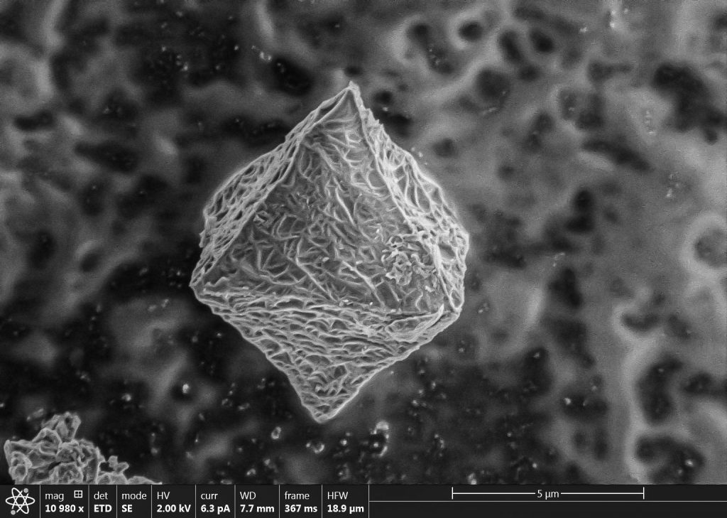
A single crystal of UiO-68 MOFs analogue with a rough surface, which makes it looks like cocoon. Microscope: SEM Teneo.

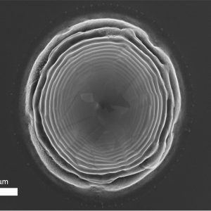
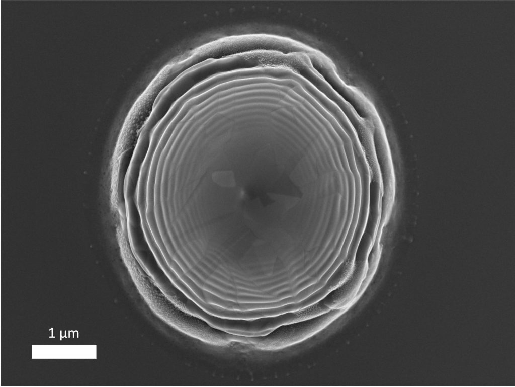
Perovskite domains featuring bend contours which are used as structural levers to mitigate strain-release and hence, formation of stacking faults. Microscope: TEM Talos.