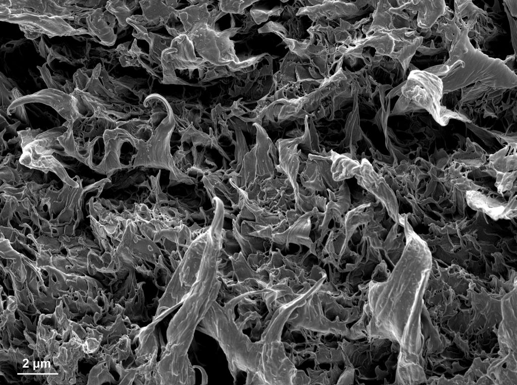
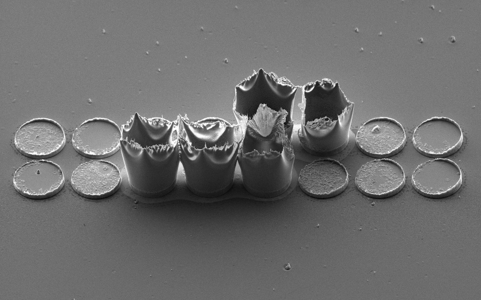
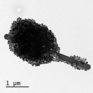
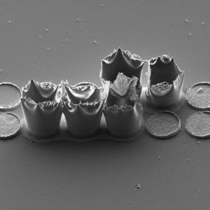
Resembling a predatory piranha bursting from the surface, this SEM image captures the dramatic fracture of a Carbon Nanotube particle. The jagged “teeth” bridging the gaping maw are actually bundles of nanotubes stretched under tension. Microscope: SEM Teneo.
Edge of the Nano-Forest, Stefano Marinoni (LMSC) September/October 2025
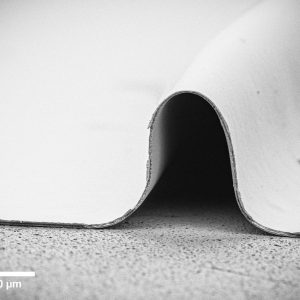
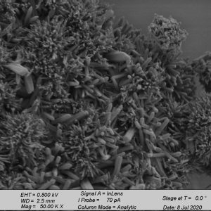
A dense, shadowed forest formed during plasma etching of a Gallium Nitride sample. The tilted SEM view captures its edge, where the trees thin out, revealing the bare substrate beneath. Microscope: SEM Merlin.
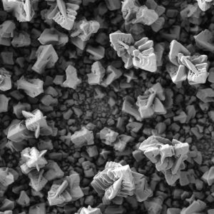
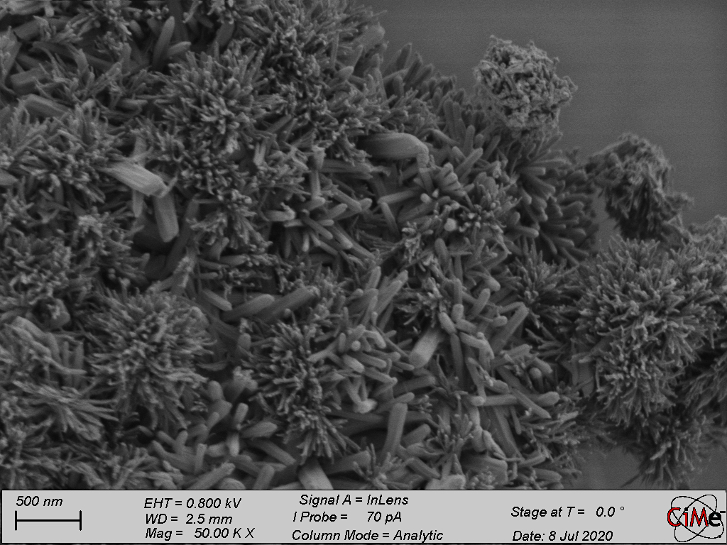
Flowers on desert, Shuqing Song (LAS) July/August 2025
This is a flower shaped ZIF crystal. Microscope: SEM Teneo.
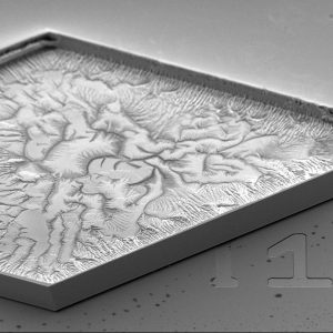
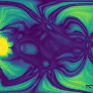
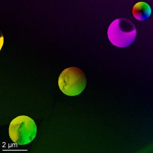
Golden Domains, Pierpaolo Ranieri (INE) May/June 2025
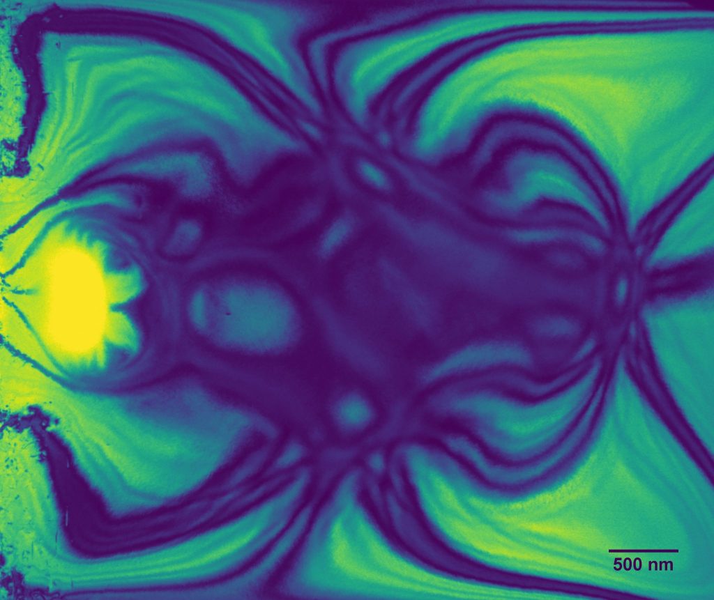
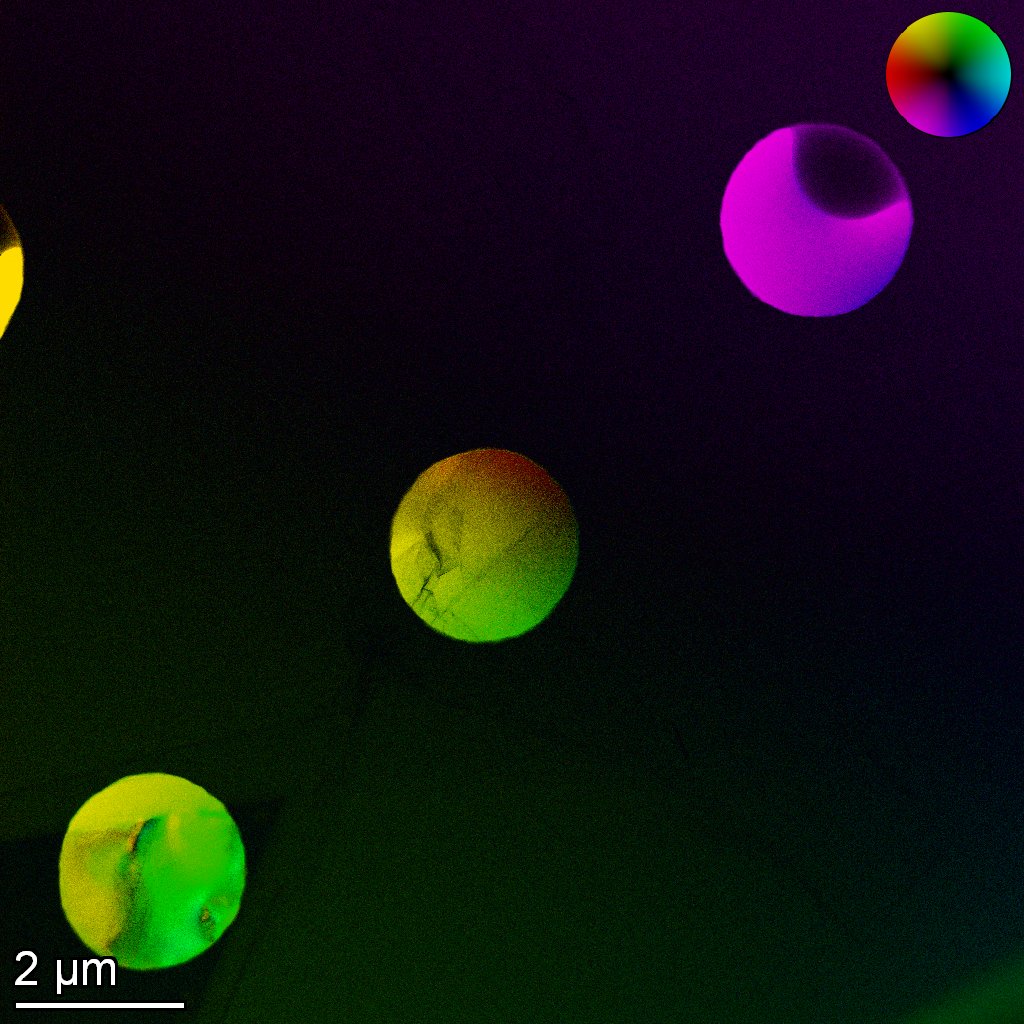
This TEM image reveals the intricate ferroelectric domain structure of a potassium niobate lamella following a thermal cycle. The pattern is false-colored in golden yellow to highlight the dynamic microstructural beauty of this functional material. Microscope: TEM Talos.
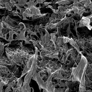
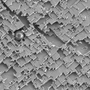
Red House on Cracked Green Earth, Qiucheng Xu (LSCI) March/April 2025
False‐colored SEM micrograph of a carbon gas diffusion electrode (GDE) surface, acquired on a field‐emission scanning electron microscope at 10 kV with the sample stage tilted ~25° to accentuate the microporous layer’s topography. The carbon surface is rendered in green and ochre tones, deep crevices appear in dark brown, and an isolated particulate feature has been false-colored bright red to resemble a small house perched above the cracked “landscape.” Microscope: SEM Gemini.
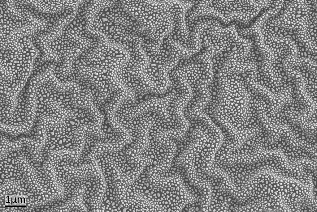
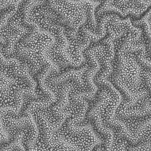
A defect in a cured transparent resist created wrinkles, forming wave-like structures. During the dewetting of a thin selenium film, droplets formed and settled on the surface, appearing to float on the waves. Microscope: SEM Gemini.
Resembling a predatory piranha bursting from the surface, this SEM image captures the dramatic fracture of a Carbon Nanotube particle. The jagged “teeth” bridging the gaping maw are actually bundles of nanotubes stretched under tension. Microscope: SEM Teneo.Edge of the Nano-Forest, Stefano Marinoni (LMSC) September/October 2025
A dense, shadowed forest formed during plasma etching of a Gallium Nitride sample. The tilted SEM view captures its edge, where the trees thin out, revealing the bare substrate beneath. Microscope: SEM Merlin.Flowers on desert, Shuqing Song (LAS) July/August 2025
This is a flower shaped ZIF crystal. Microscope: SEM Teneo.Golden Domains, Pierpaolo Ranieri (INE) May/June 2025
This TEM image reveals the intricate ferroelectric domain structure of a potassium niobate lamella following a thermal cycle. The pattern is false-colored in golden yellow to highlight the dynamic microstructural beauty of this functional material. Microscope: TEM Talos.Red House on Cracked Green Earth, Qiucheng Xu (LSCI) March/April 2025
False‐colored SEM micrograph of a carbon gas diffusion electrode (GDE) surface, acquired on a field‐emission scanning electron microscope at 10 kV with the sample stage tilted ~25° to accentuate the microporous layer’s topography. The carbon surface is rendered in green and ochre tones, deep crevices appear in dark brown, and an isolated particulate feature has been false-colored bright red to resemble a small house perched above the cracked “landscape.” Microscope: SEM Gemini.Sea-lenium Waves_Laurène Tribolet (FIMAP) January/February 2025
A defect in a cured transparent resist created wrinkles, forming wave-like structures. During the dewetting of a thin selenium film, droplets formed and settled on the surface, appearing to float on the waves. Microscope: SEM Gemini.
“Seeing is believing.” This freely suspended structure was fabricated by two-photon lithography (TPL), followed by atomic layer deposition (ALD) of Ni on a SiN membrane frame without a membrane. The free-form design achieved by TPL and the 3D conformal coating by ALD are being seen. Microscope: SEM Gemini.
Le masque du fantôme triste, Ariana Serban (LSCI) September/October 2024
Ni foam support used in water splitting that looks like a sad, hanging ghost. Microscope: SEM Gemini.
Dendritic Algae Reef, Bouchez Arthur (LIMNO) July/August 2024
Salt dendrites with polymer nanoparticles. Observed by TEM. Microscope: TEM Talos.
Goldfish with human face, Shuqing Song (LAS) May/June 2024
The image looks quite like a goldfish with a human face. Microscope: SEM Teneo.
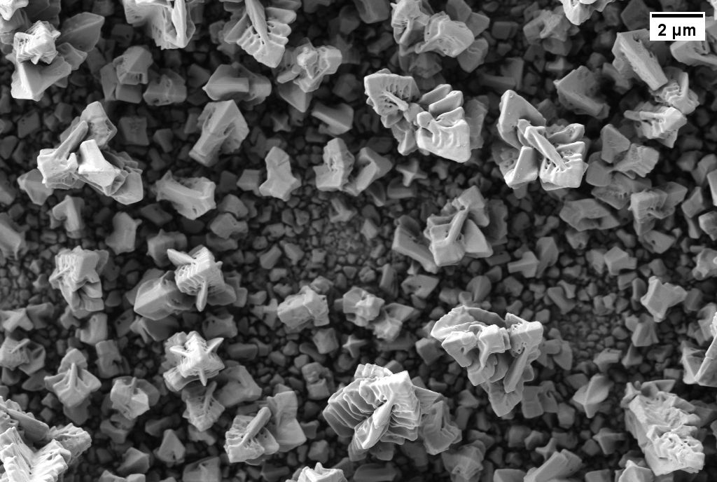
Microscopic Bloom – A Rose Unveiled, Thi Ha My Pham (LMER) March/April 2024
Gas Diffusion Electrode after electrochemical CO2 reduction. Microscope: SEM Teneo.
An undesirable air bubble created a hole in a flat substrate, which turned into a dark moon hiding the sun in a starry sky after the dewetting of a thin selenium film. Microscope: SEM Gemini.
“Seeing is believing.” This freely suspended structure was fabricated by two-photon lithography (TPL), followed by atomic layer deposition (ALD) of Ni on a SiN membrane frame without a membrane. The free-form design achieved by TPL and the 3D conformal coating by ALD are being seen. Microscope: SEM Gemini.Le masque du fantôme triste, Ariana Serban (LSCI) September/October 2024
Ni foam support used in water splitting that looks like a sad, hanging ghost. Microscope: SEM Gemini.Dendritic Algae Reef, Bouchez Arthur (LIMNO) July/August 2024
Salt dendrites with polymer nanoparticles. Observed by TEM. Microscope: TEM Talos.Goldfish with human face, Shuqing Song (LAS) May/June 2024
The image looks quite like a goldfish with a human face. Microscope: SEM Teneo.Microscopic Bloom – A Rose Unveiled, Thi Ha My Pham (LMER) March/April 2024
Gas Diffusion Electrode after electrochemical CO2 reduction. Microscope: SEM Teneo.Lunar eclipse, Laurène Tribolet (FIMAP) January/February 2024
An undesirable air bubble created a hole in a flat substrate, which turned into a dark moon hiding the sun in a starry sky after the dewetting of a thin selenium film. Microscope: SEM Gemini.
Dendritic growth of copper during electrodeposition resembling blooming of flowers. Microscope: SEM/FIB CrossBeam.
Roaring Fire, Yan Meng (FIMAP) July/August 2023
Polyvinylidene Difluoride (PVDF) polymer thin film after thermally drawing. This soft and plastic film under microscopy looks like fire burning with flames. Microscope: SEM Gemini.
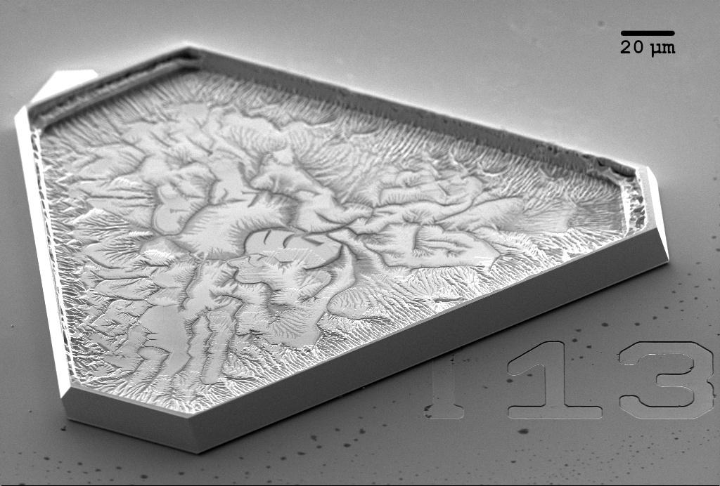
SEM image of a chemically synthesized gold crystalline flake exhibiting a fractal-shaped defect, reminiscent of von Koch fractal. Perhaps, the crystal was unlucky to get a defect during the growth as it was labelled with the unlucky number 13? Microscope: SEM/FIB CrossBeam.
For 2023 St-Patrick’s day: an Irish fiddle made of the stacking of Cu cubes and few Cu rods. Microscope: TEM Spirit.
Tunnel to nowhere, Cédric Van Goethem (LAS) January/February 2023
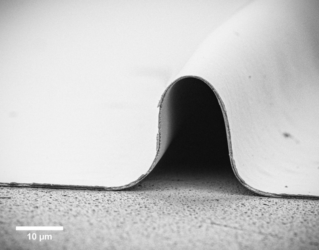
Unwanted wrinkle in a porous nanocrystalline graphene membrane. Microscope: SEM Teneo.
Selenium Valley, Laurène Tribolet, Eva Vogt (FIMAP) November/December 2023
Thin layer of selenium dewetted into beautifully spread droplets on winding resist valleys resulting from unwanted buckling. Microscope: SEM Gemini.Dendretic flowers – Ayush Khurana (TIC) September/October 2023
Dendritic growth of copper during electrodeposition resembling blooming of flowers. Microscope: SEM/FIB CrossBeam.Roaring Fire, Yan Meng (FIMAP) July/August 2023
Polyvinylidene Difluoride (PVDF) polymer thin film after thermally drawing. This soft and plastic film under microscopy looks like fire burning with flames. Microscope: SEM Gemini.von Koch’s goldflake #I13, Sergejs Boroviks (STI – NAM) May/June 2023
SEM image of a chemically synthesized gold crystalline flake exhibiting a fractal-shaped defect, reminiscent of von Koch fractal. Perhaps, the crystal was unlucky to get a defect during the growth as it was labelled with the unlucky number 13? Microscope: SEM/FIB CrossBeam.Nano Irish Fiddle, Ludovic Zaza (LNCE) March/April 2023
For 2023 St-Patrick’s day: an Irish fiddle made of the stacking of Cu cubes and few Cu rods. Microscope: TEM Spirit.Tunnel to nowhere, Cédric Van Goethem (LAS) January/February 2023
Unwanted wrinkle in a porous nanocrystalline graphene membrane. Microscope: SEM Teneo.
Nothing better than a nano ice cream during a hot summer afternoon. Composed of randomly distributed Cu nanocrystals around a Cu rod. Microscope: TEM Tecnai G2 Spirit Twin (Sion).
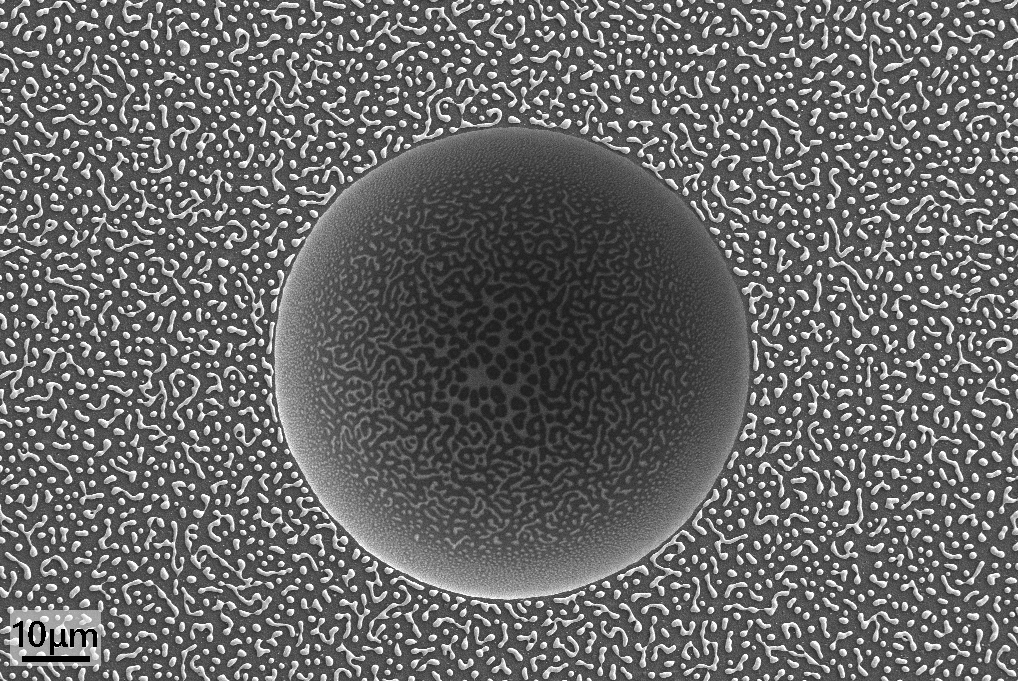
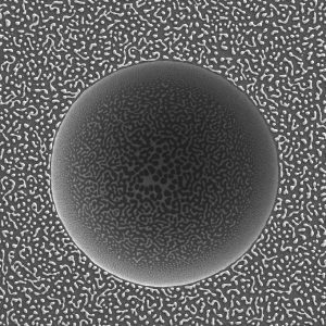
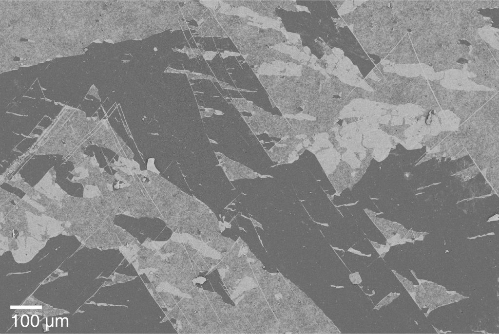
Geometries, Alejandra Slagter (LMM) May/June 2022
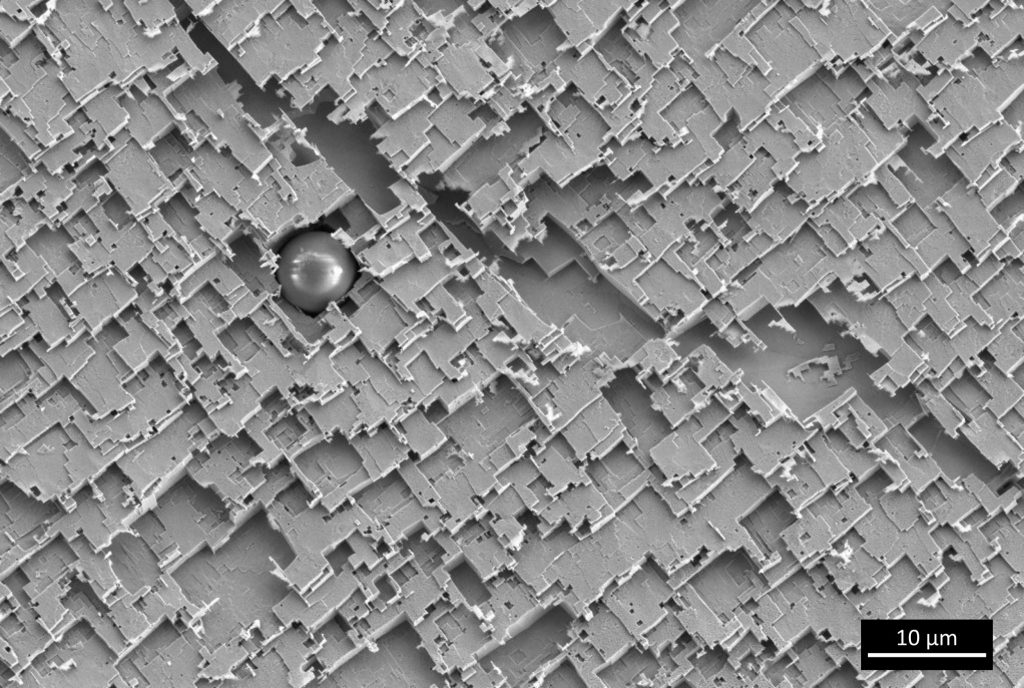
A spherical inclusion sitting on a chemically etched iron surface. Microscope: Gemini SEM
Ge sea monsters, Andrea Giunto (LMSC) March/April 2022
Germanium monsters arose at the bottom of a Silicon ocean from a Ion Beam Etching gone wrong. They are waiting with their gaping jaws for the next pray passing by. Microscope: SEM/FIB NVision.
Cement coral, Andrea Teixeira (LMC) January/February 2022
Calcium silicate hydrate (C-S-H) and ettringite coral in a cement sample after one day of hydration. Microscope: SEM Merlin.
Freeze-dried polymeric core shell particle. Microscope: SEM Gemini.Spider TEM, Pierpaolo Ranieri (INE) September/October 2022
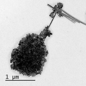
Potassium niobate lamella prepared using focused ion beam. As an effect of the bend contours a spider appears on the surface. Microscope: TEM Talos.Nano Ice-Cream, Ludovic Zaza (LNCE) July/August 2022
Nothing better than a nano ice cream during a hot summer afternoon. Composed of randomly distributed Cu nanocrystals around a Cu rod. Microscope: TEM Tecnai G2 Spirit Twin (Sion).Geometries, Alejandra Slagter (LMM) May/June 2022
A spherical inclusion sitting on a chemically etched iron surface. Microscope: Gemini SEMGe sea monsters, Andrea Giunto (LMSC) March/April 2022
Germanium monsters arose at the bottom of a Silicon ocean from a Ion Beam Etching gone wrong. They are waiting with their gaping jaws for the next pray passing by. Microscope: SEM/FIB NVision.Cement coral, Andrea Teixeira (LMC) January/February 2022
Calcium silicate hydrate (C-S-H) and ettringite coral in a cement sample after one day of hydration. Microscope: SEM Merlin.
Solar system model, Mukesh Tripathi (LANES) November/December 2021
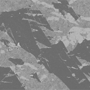
Differential phase contrast (DPC) imaging of freestanding 2D materials with thickness variations. Microscope: TEM Themis
3D nano-ant walking in TiAl alloy – Gulnaz Ganeeva (LSME) September/October 2021
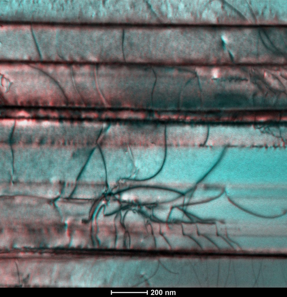
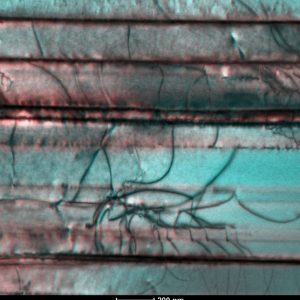
Anaglyph made from tilt-less stereo-pair STEM images of dislocations in TiAl alloy, stereo-angle approximately 2 degrees. Best viewed with 3D glasses. Microscope: TEM Osiris.
Micrometer Mountains of Valais, Luis Francisco Villalobos (LAS) May/June 2021
A failed yet beautiful attempt towards the optimization of poly(triazine imide) membranes. We deposited single-layer poly(triazine imide)nanosheets on a porous alumina support. Microscope: SEM Teneo.
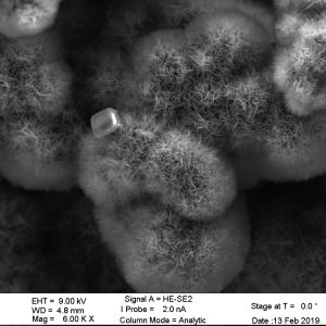
Formation of silico-alkali-calcareous “hedgehogs” in concrete immersed for 3 months in an alkaline solution. Microscope: SEM Merlin.
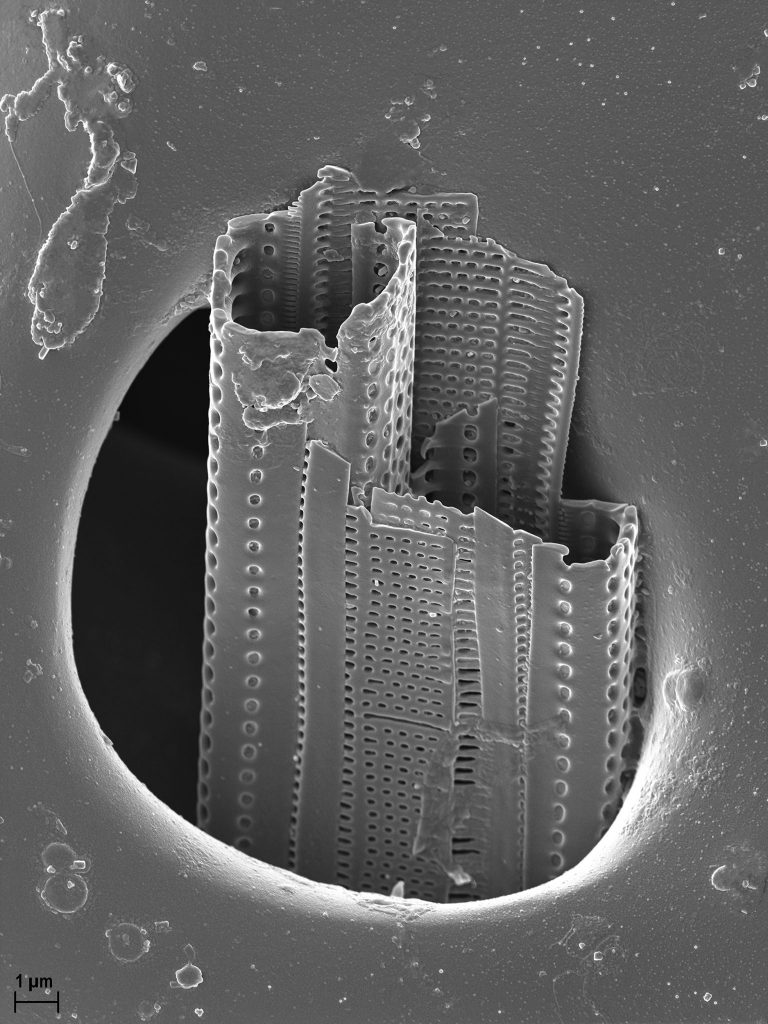
Seascraper, France Bourely (LNMC) January/February 2021
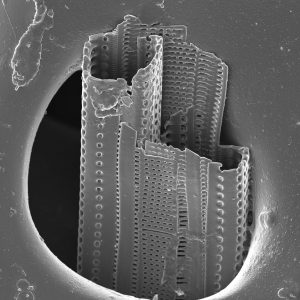
Phytoplancton emerging from a pore on the skeleton of the mediterranean sea urchin Paracentrotus lividus. The function of the many interconnected pores is to allow the passage of adhesive tube feet, mainly for locomotion. This endoskeleton, enclosed by an epidermis, and spines, is called a test. It is famous for its beautiful symmetry, and is mostly made of calcite and magnesite. Its shape is effective at distributing stress evenly over the surface. Marine echinoderms can be studied for their numerous biomimetic applications. Gold coated, and photographed at 2kv on a Gemini-SEM 300.
Solar system model, Mukesh Tripathi (LANES) November/December 2021
Differential phase contrast (DPC) imaging of freestanding 2D materials with thickness variations. Microscope: TEM Themis3D nano-ant walking in TiAl alloy – Gulnaz Ganeeva (LSME) September/October 2021
Anaglyph made from tilt-less stereo-pair STEM images of dislocations in TiAl alloy, stereo-angle approximately 2 degrees. Best viewed with 3D glasses. Microscope: TEM Osiris.Micrometer Mountains of Valais, Luis Francisco Villalobos (LAS) May/June 2021
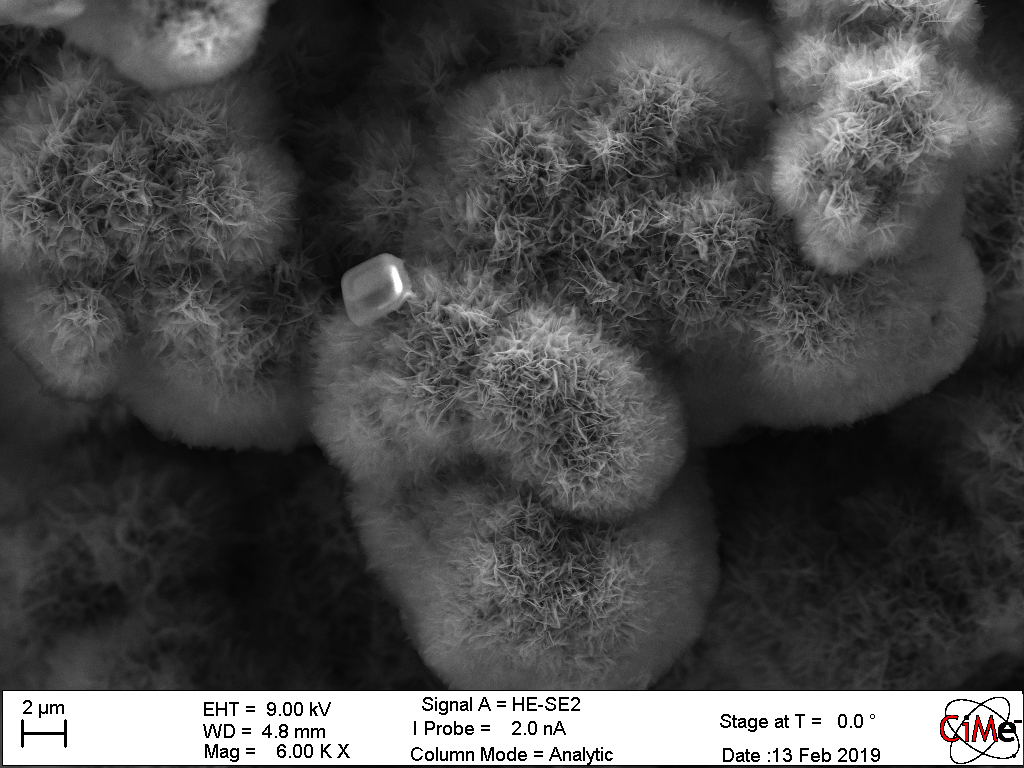
A failed yet beautiful attempt towards the optimization of poly(triazine imide) membranes. We deposited single-layer poly(triazine imide)nanosheets on a porous alumina support. Microscope: SEM Teneo. Furry Friends – Solene Barbotin-Albinski (LMC) March/April 2021
Formation of silico-alkali-calcareous “hedgehogs” in concrete immersed for 3 months in an alkaline solution. Microscope: SEM Merlin.Seascraper, France Bourely (LNMC) January/February 2021
Phytoplancton emerging from a pore on the skeleton of the mediterranean sea urchin Paracentrotus lividus. The function of the many interconnected pores is to allow the passage of adhesive tube feet, mainly for locomotion. This endoskeleton, enclosed by an epidermis, and spines, is called a test. It is famous for its beautiful symmetry, and is mostly made of calcite and magnesite. Its shape is effective at distributing stress evenly over the surface. Marine echinoderms can be studied for their numerous biomimetic applications. Gold coated, and photographed at 2kv on a Gemini-SEM 300.