Scanning TEM (STEM)
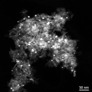
The condenser lens system of the microscope is used to focus a small electron probe on the sample. Scan coils then raster the probe across the sample (similarly to SEM). While a central bright-field detector may exist, images from an annular dark field (ADF) detector are usually of more interest. Such ADF images, formed from electrons incoherently scattered at high angles, have image intensities dependent on sample thickness and atomic number (“Z contrast” images). These images can therefore give good compositional contrast, for instance of heavy catalyst particles on light supports, or of multilayers made from different materials. When combined with EDXS or EELS, STEM becomes a powerful technique for analysing compositional variations on nanometric to micrometric scales.