Bright field (BF)
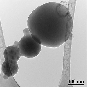
Imaging of the sample under broad electron beam illumination, and with an objective aperture selecting only undiffracted electrons for the image formation (e.g. to improve image contrast and focusing). BF images contain mass-thickness and diffraction contrasts. In crystalline specimens, one may observe thickness fringes, bend contours, grain boundaries, crystalline defects such as dislocations, twins, or stacking faults, and second phases. Nevertheless, general BF imaging requires minimal set up, and is, for instance, useful for specimen inspection, and for imaging size and morphology of nanostructures such as nanotubes or nanoparticles. BF imaging is also the main technique for observation of biological specimens, which are typically stained with heavy metals to give mass contrast.