The BIOP hosts over 20 light microscopes with imaging modalities ranging from stereomicroscopy to super-resolution. Below you will find a (non-exhaustive) list of the techniques available on the platform.
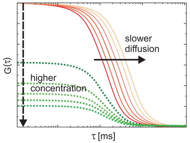
FCS is a correlation analysis of fluctuation of the fluorescence intensity. The analysis provides parameters of the physics under the fluctuations. One of the interesting applications of this is an analysis of the concentration fluctuations of fluorescent particles (molecules) in solution.
Available Microscopes: Zeiss LSM710
Contact: Arne Seitz
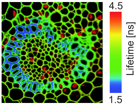
Fluorescence Lifetime Imaging Microscopy (FLIM) uses dedicated hardware that enables the calculation of the time between a fluorophore being excited and when a photon is released, called the Lifetime). They are often used in FRET (Förster resonance energy transfer) experiments.
Available Microscopes: Leica SP8 FLIM
Contact: Nicolas Chiaruttini, Romain Guiet
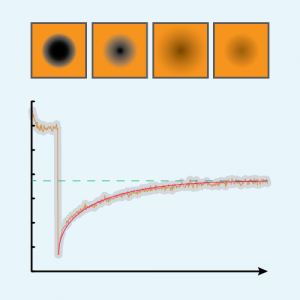
FRAP-like experiments are used to quantify the mobility of proteins in cells or tissues. They require high laser power and scanners to bleach rapidly and precisely the fluorophores of interest.
Available Microscopes: Leica SP8 IN1, Leica SP8 UP1, Leica SP8 UP2, Zeiss LSM 710
Contact: José Artacho, Nicolas Chiaruttini
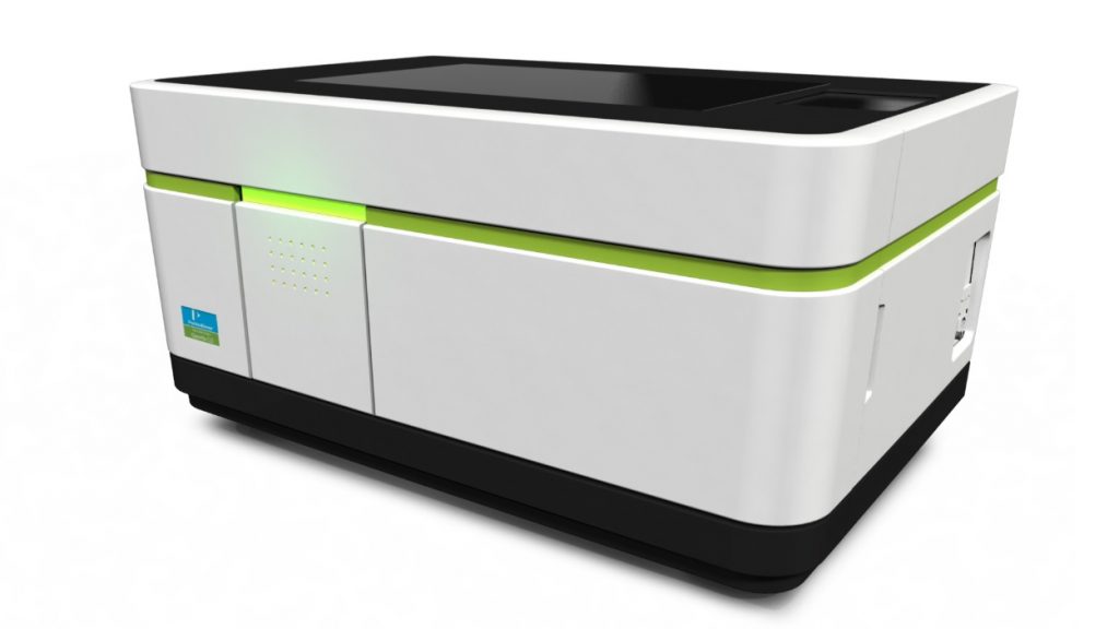
High throughput imaging microscopes are usually dedicated systems built to handle well-plates, boast excellent stability for long-term imaging and are usually hardware optimized for fast acquisitions.
Available Microscopes: PerkinElmer Operetta
Contact: José Artacho, Nicolas Chiaruttini
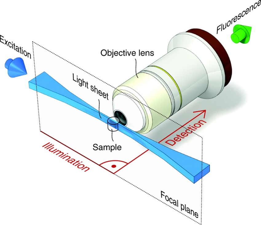
Lightsheet, or Selective Plane Illumination Microscopy (SPIM), allows for fast gentle fluorescence imaging with optical slicing and multi-angle observation of both live and cleared samples.
Available Microscopes: Zeiss Lightsheet Z1
Contact: Nicolas Chiaruttini
Live imaging systems are usually inverted microscopes with temperature, humidity and CO2 control in order to host delivate live samples for long-term imaging.
Available Microscopes: LSM710, Zeiss Lightsheet Z1, Leica SP8-FLIM, Leica SP8 STED 3X, Visitron CSU W1, PerkinElmer Operetta.
Microdissection microscopes utilize pulsed UV lasers to perform localized micro-ablations in tissue or within living cells. This can be used for isolating fresh tissue samples for RT-PCR or altering biological processes in cells/organisms in order to study their influence.
Available Microscopes: Zeiss PALM
Contact: José Artacho
Super-resolution techniques have gained momentum over the last few years, pushing the limits of light microscopy beyond the classical diffraction limit.
At the BIOP, you can perform Structured Illumination (SIM), Stimulated Emission Depletion (STED) and Stochastic Optical Reconstruction (STORM).
Available Microscopes: Nikon Crest, Abberior Mirava, Leica SP8 STED 3X,
Contact STED: José Artacho, Nicolas Chiaruttini
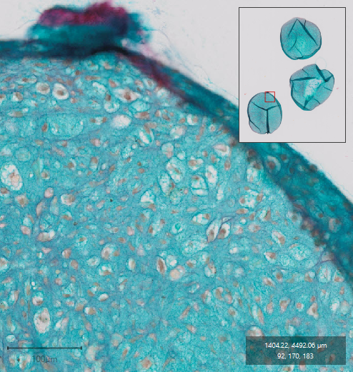
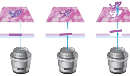
Whole Slide Scanners allow the acquisition of large areas in minimum time and high magnification (up to 40x). Data can then be viewed and processed in dedicated software, such as QuPath.
The BIOP offers two fully automated whole slide scanners with 100-slide capacity. Imaging modalities include brightfield and fluorescence. For Confocal tile scanning, our new Leica SP8 Microscopes with Navigator are a viable option as well.
Available Microscopes: Olympus Slide Scanner 2, Olympus Slide Scanner 3 and Olympus Slide Scanner 4, Leica SP8 IN1, Leica SP8 UP1, Leica SP8 UP2.
Contact: José Artacho, Nicolas Chiaruttini, Rémy Dornier