Microsurgery
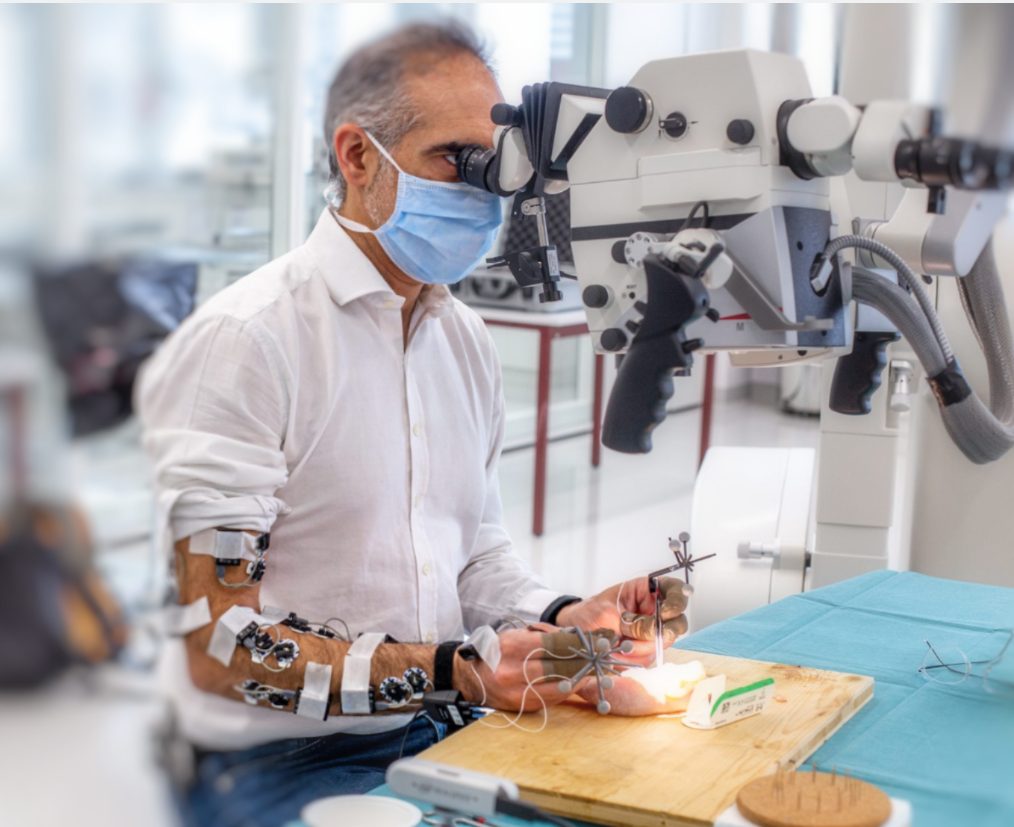
Training model for anastomosis. The aim is to suture back a 1mm large chicken wing artery. The surgeon uses position trackers on the tools and wears EMG sensors to record muscles activities and a finger TPS (Tactile Pressure Sensing) system. Credit : SFITS
Motivation
Introduction
Microsurgery is a type of surgery that uses powerful microscopes, specialized precision tools and various operating techniques. It is primarily used to anastomose small (1-2 mm) blood vessels i.e. to join two vessels together so that the blood can flow though and to coapt nerves.
Microvascular and microneural coaption are used for complex repair of human tissue, organ transplantations and revascularization of the brain.
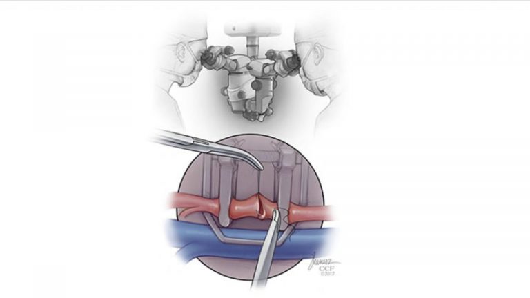
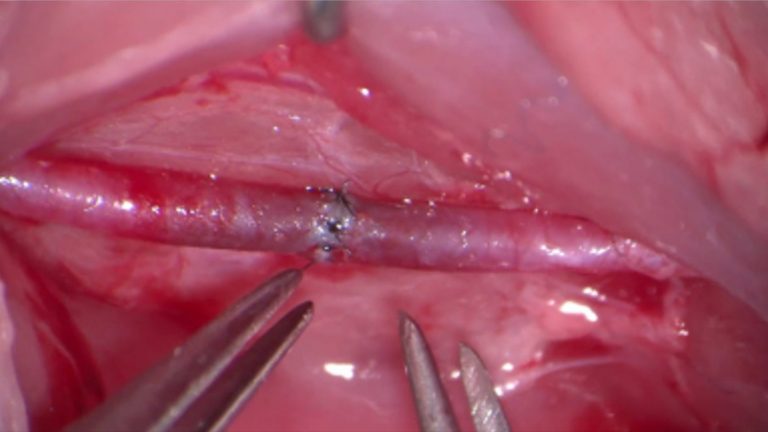
Image: Example of a microsurgical operation with an end-to-end microvascular anastomosis [1].
Problem
Microsurgery involves a unique set of surgical principles and is technically much more demanding than routine surgical practice. Operating on vessels with such small calibers under microscopic vision offers little or no haptic feedback, a limited dimensional perspective, and requires superior dexterity. The margin of error in microvascular surgery is between 1/50 and 1/100 mm and every small mistake increases the probability of vessel occlusion exponentially. Consequently, it demands very fine hand movements in combination with stereoscopic vision and visuospatial skills
Goal
Teaching and assessment of skill acquisition in microsurgery is tricky as there are not clear and objective standards. It relies on subjective opinions of senior faculty. However, optimizing the training of future microsurgeons is of great importance and includes objective assessment of skills, learning curves, and value of various training models.
The goals of the proposed project is to add scientific knowledge on how hand position, force application and movements change during the surgeon’s trainings and to advance clinical teaching by developing an objective evaluation tool for the assessment of microsurgical competence.


Acknowledgments
The goals will be achieved through an interdisciplinary collaboration between micro-neurosurgeons at the University Hospital of Geneva (HUG), the University of Medicine and Pharmacy (UMF) of Cluj-Napoca and experts in signal processing and human modeling at EPFL.
LASA
The project is conducted by the Learning Algorithms and Systems Laboratory (LASA) at the Swiss Federal Institute of Technology Lausanne (EPFL).

SFITS
This project is in collaboration with micro-surgeons from the SFITS at HUG (Hôpitaux Universitaires de Genève).
UMF Cluj-Napoca
This project is in collaboration with micro-surgeons from the University of Medicine and Pharmacy (UMF) of Cluj-Napoca

Funded by
SNSF
This project is supported by the Swiss National Science Foundation. Project number 325230_204694.
More

This website follows EPFL Regulations on Data Protection
Sources
1. https://my.clevelandclinic.org/departments/dermatology-plastic-surgery/medical-professionals/microsurgery-training-courses
2. Chan, W. Y., Matteucci, P. & Southern, S. J. Validation of microsurgical models in microsurgery training and competence: a review. Microsurgery 27, 494-499, doi:10.1002/micr.20393 (2007).