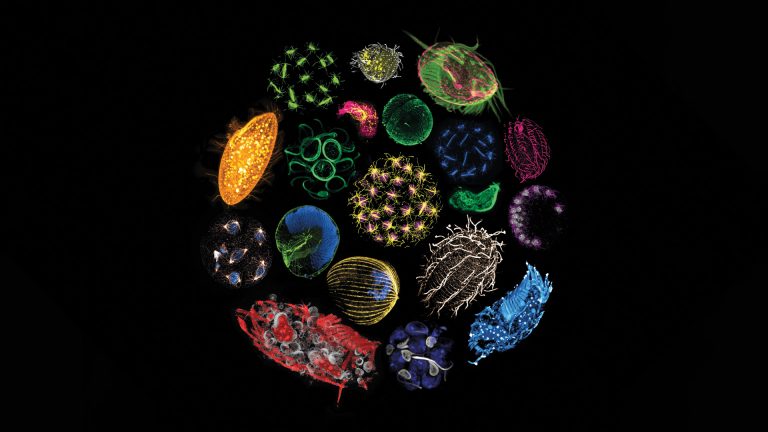
At the intersection of life sciences, imaging and art, VOIR EST SAVOIR celebrates the richness of scientific imaging and the beauty of sciences over many scales
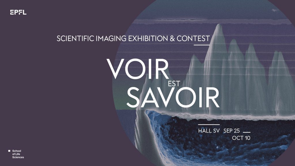
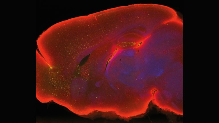
Witness the front of the mouse brain, featuring the prefrontal cortex. Notice the yellow neurons responsible for traumatic memory formation, alongside the blue cell nuclei. Through TRAP transgenic mice and fluorescence microscopy, we explore memory pathways, unraveling anatomical, physiological, and molecular insights. This project unveils the intricate workings of memory formation, bridging art and science to ignite curiosity and appreciation for the complexities of the memory formation.
Myroslava Volosko, Intern at the Gräff Lab.
I am captivated by the beauty and mysteries of the brain. Neuroscience drives my passion to uncover its enigmatic depths.
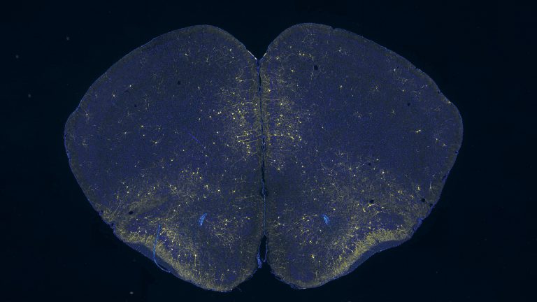
Immunofluorescently stained organoids: Miniature tumor models revealing cancer treatment insights. These organoids mimic tumors, aiding research on treatment response. The vibrant colors represent specific biomarkers, guiding the study of cancer pathways and therapy efficacy. Organoids offer personalized insights, accelerating drug screening and reducing reliance on animals models. Advancing cancer research, they optimize treatment for better patient outcomes.
Laure Garnier, Organoid Culture & 3D Models Expert at the Bioengineering & Organoids platform.
I am bridging basic science and practice for clinical applications, advancing diagnostics, development.
Voir Est Savoir 2023
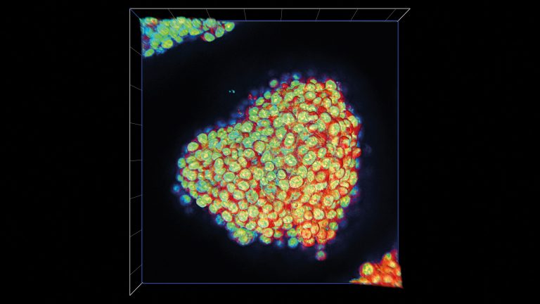
Cerebral functions rely on diverse cells, including astrocytes, the most abundant. Astrocytes play key neurological roles with their star-shaped architecture (cyan). Briefly, multiple branches extend from the soma and radially ramify into finer ones. Some branches enlarge at their extremities, called endfoot, to surround blood vessels (red) and form the blood-brain barrier. Our project aims to better characterize these cells and endfeet anatomically and molecularly.
Maximum intensity projection image. Microscope Leica SP8 Upright. Objective 40x glycerol. Pixel size: 86nm;Z-step:180 nm.
Charlotte Lorin, Postdoc at the Blue Brain Project.
I am still, after many years in science, amazed by cutting-edge technologies like microscopy, giving the privilege to visualize the biology wonders.
Ultrastructural diversity of distinct plankton species revealed by expansion microscopy.
Omaya Dudin, Group leader at the Dudin Lab.
We are interested in the cell & developmental biology of protists, with a main focus on Ichthyosporeans, which are close animal relatives exhibiting animal-like development.
Bronze Medal
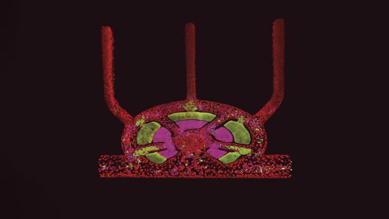
Lymph nodes are key to the adaptive immune response. Composed of specialised domains for B and T cells within a lymphoid vascular framework, the lymph node is a complex organ. So complex, it has been impossible to date to accurately model the structure in vitro. Here we show the first multi-cell, spatially precise lymph node on a chip, with its lymphatic endothelium (red), B lymphocytes (green), and T lymphocytes (magenta). Notice the striking resemblance to the iconic Millennium Falcon cockpit?
Jakob J. Langer & L. Francisco Lorenzo Martin, researchers at the Lutolf Lab.
We are driven by our passion for developing sophisticated mini-organs on-a-chip that capture human physiology in all its detail.
Silver Medal
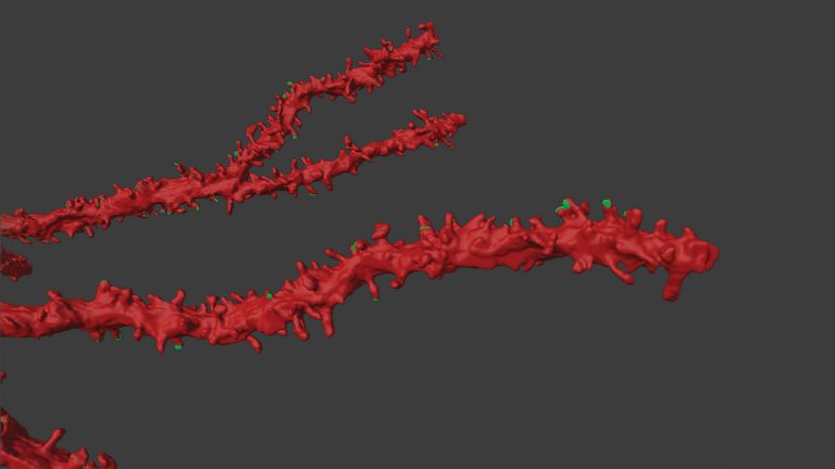
The picture shows a three-dimensional reconstruction of dendritic segments (in red) and synapses (in green) of a neuron involved in the storage of memory. These neurons are called engrams, they are activated during learning and are thought to store the mnemonic trace. In my project we want to investigate and understand where remote memory is stored. To look at the deep ultrastructural level of engrams we used transmission electron microscopy and we exported the neuron in 3D.
Isabella Tarulli, PhD student at the Gräff Lab.
I have a strong interest in molecular neurobiology. I am fascinated on how chromatin and epigenetic mechanisms are involved in neurobiological processes, such as memory.
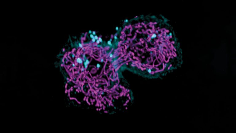
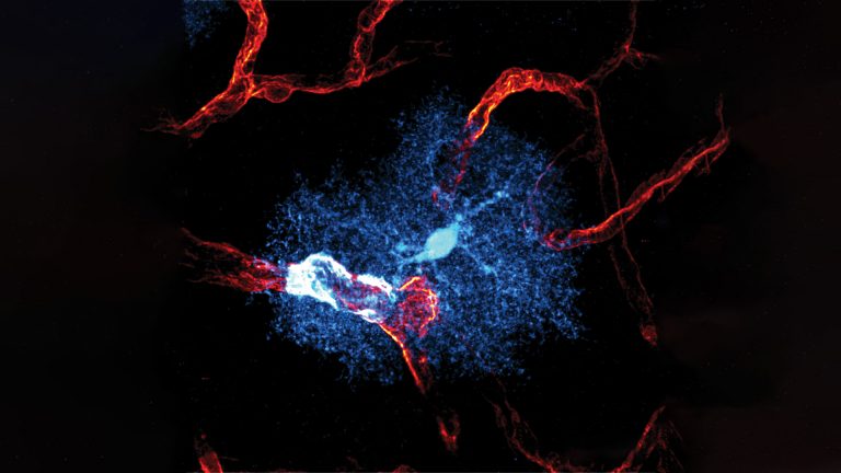
While investigating how mitochondria (magenta) move through a cell and share material and information between subpopulations of a cell I stumbled across a cell undergoing cell division. In this snapshot of a movie you can see the cell slowly constricting the division site and the mitochondria being equally distributed among the daughter cells. Cortical actin (cyan) is also visible and particularly bright at the constriction site. This image was acquired on the lattice lightsheet at the BIOP.
Julius Winter, PhD student at the Manley Lab.
I am fascinated with cellular processes occurring at the microscopic scale.
I aim to dissect the physical substrate of memory trace termed as engram. I performed brain clearing which renders the rodent brain transparent, and imaged whole brain with light sheet microscope. Immunostaining against cfos in red and epigenetic mark in green was applied to visualize how memory is formed and stored both at brain network and epigenetic state.
Kwok Yui Reymond Yip (Tony), PhD student at the Gräff Lab.
I investigate if memory engram cells are epigenetically defined at brain-wide level.

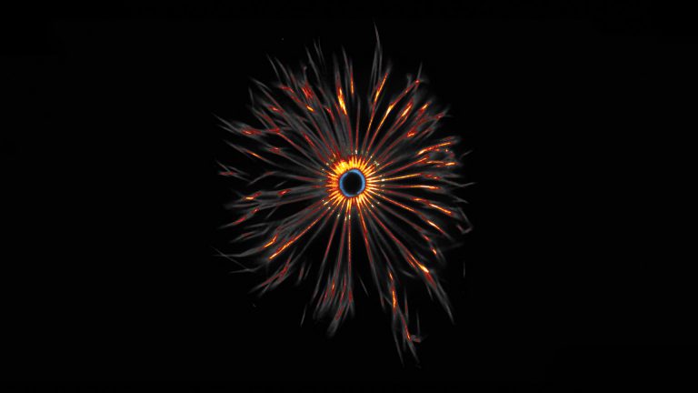
Telomeres, nucleoprotein structures at the end of chromosomes and composed of repetitive nucleotide sequences, were detected by FISH probes. Red indicates lagging strands and yellow indicates leading strands. These shining stars in the human galaxy were aligned like the milky way.
Suna In, Postdoc at the Lingner Lab.
My research aims to investigate what triggers telomeric FISH signals on metaphase chromosomes to become smeary or doubled, termed as “fragile”.
A top view of the unicellular organism, Trichonympha agilis, decorated by thousands of flagella used to navigate the dense environment of the termite hindgut. Each is templated by one structure: the centriole. To investigate the molecular identity of its components, we use “Expansion Microscopy” to physically separate molecules in space to uncover details never before seen by optical microscopy.
Alana Dastous, PhD student at the Gönczy Lab.
To better understand centriole biogenesis, I aim to uncover the molecular identity of core centriole components and to decipher their role in this process.

Progressive degeneration of neurons expressing human Tau. Alterations of the microtubule associated protein Tau are associated with many human neurodegenerative disorders. Employing adult onset expression of human Tau in Drosophila Dorsal Cluster (DC) neurons, we could observe and quantitate with single neuron resolution progressive neurodegeneration and axon loss within the context of an intact whole brain.
Left side: control DC neuron axons Right side: DB axons expressing human Tau degenerate.
Jamshid Asadzadeh Postdoctoral Scientist in the McCabe lab
I was a Postdoctoral Scientist in the McCabe lab in the EPFL Brain Mind Institute until 2021. I am now a Clinical Data Manager at Clario.
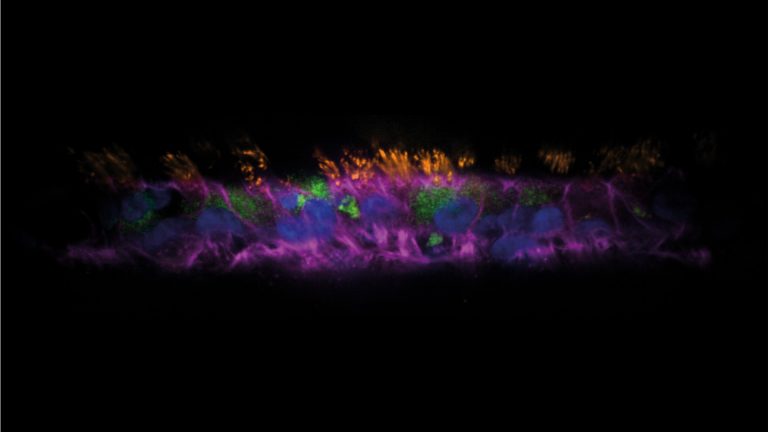
This shows a human airway organoid called AirGel. It comprises relevant cell types like mucus-secreting (green) and ciliated (orange) cells. When cilia beat, they generate a flow that propels mucus out of the respiratory tract, along with any pathogen trapped in it. As they allow to monitor how bacteria interact with airway mucus dynamically, AirGels are valuable tools for research on respiratory infections, hopefully opening the way for new strategies to face the upcoming antibiotic crisis.
Tamara Rossy, PhD student at the Persat Lab.
I was a PhD student in the Persat Lab until May 2023. I focused on studying bacteria in realistic environments – such as AirGels to mimic the human airways. I am now a postdoctoral researcher at MIT.
The image represents 3D lung tumor spheroids, grown in flipped-well hanging-drop conditions. The aim of the project is to assess how innate immunity can, counterintuitively, help tumor progression, such as helping the cells survive and stick together.
Anita Bodac, PhD student at the Radtke and Meylan Labs.
My aim is to find a way to target immune cells associated with poor patient survival in lung cancer.
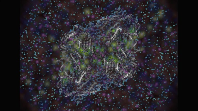
Embark on a mesmerizing journey into the cryo-electron microscopic universe of macromolecules. Imagine a galaxy of 160Å wide, and you wind up with the majestic β-Galactosidase. A cosmic dance of knowledge unfolds at atomic resoltion, helping scientists to reveal its mysteries. Here, in an artistic’s way, we unveil the saga of life’s building blocks.
Yoan Duhoo, scientist at the Protein Production Platform (PTPSP), specialized in cryo-electron microscopy and protein structure characterization.
Above all, I have a fondness for venomous animals!
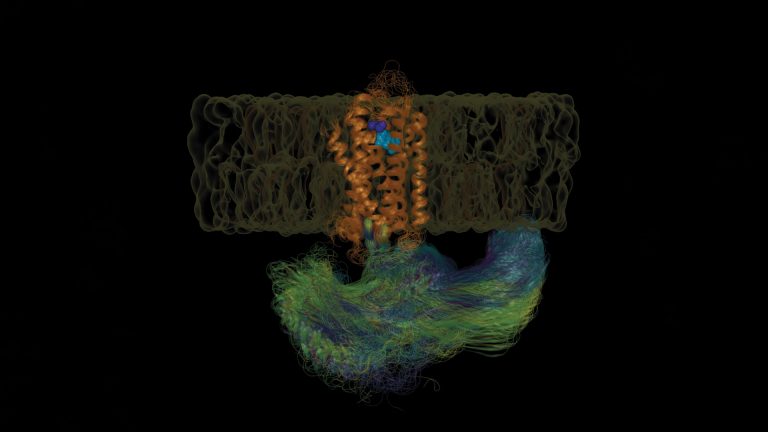
How does a psychedelic molecule, such as LSD, trigger such an intense experience for us? Shown is the simulated motion of membrane bound serotonin 2B (orange) receptor in response to LSD (cyan/purple) and binding an intracellular partner (yellow thru blue), which will signal inside the cell. The purple part of LSD is critical for its hallucinogenic effect. The overarching project is a study of long-range communication in G-protein coupled receptors, of which serotonin receptors are a part.
Mahdi Hijazi, PhD student at the Barth Lab.
I am interested in giving life to static protein structures using molecular simulations and uncovering how proteins communicate.
Winner 2023
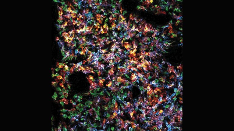
The image portrays stem cells in cell culture stained for glycosphingolipids, which are present in the plasma membrane. Being very diverse and heterogenous, the stem cell population mirrors human population. Each subject, a cell or a human, stands as a masterpiece of intricacy and singularity. Yet, when united in purpose, they bestow upon us a mosaic of unparallel splendour, the most exquisite portrait imaginable, a testament to the harmonious dance of life’s forms.
Nika Gorsek, PhD student at the D’Angelo Lab.
I would like to unravel the lipid heterogeneity of stem cells on the single cell level during development.
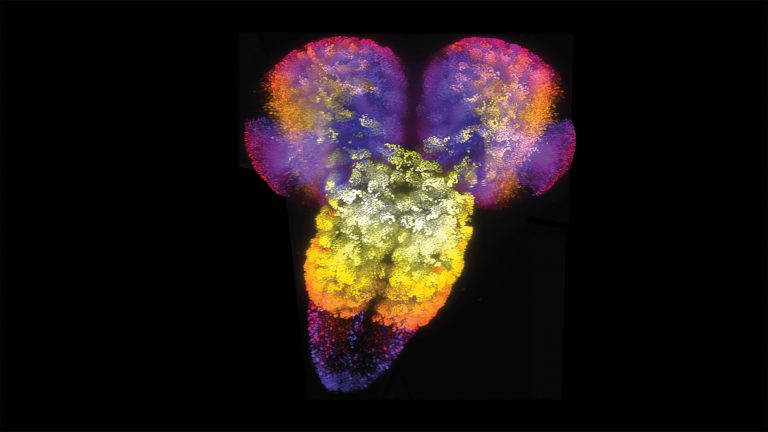
Using genetic tools designed for computer-assisted science, the exact number of cells in the entire Drosophila third instar larval central nervous system (CNS) was quantitated, revealing less functional neurons and more glia than previously predicted, plus that female larva have 9.75% more neurons than males. The image shows a light sheet image of the entire Drosophila larval CNS, with neuronal stem cells labelled by inscuteable Gal4 and coloured based upon Z-position.
Wei Jiao, Postdoc in McCabe lab.
I was a postdoctoral scientist in the McCabe lab until 2022. I am now a Junior Lecturer at the University of Lausanne. The McCabe lab is interested in motor neuroscience and motor system disease.