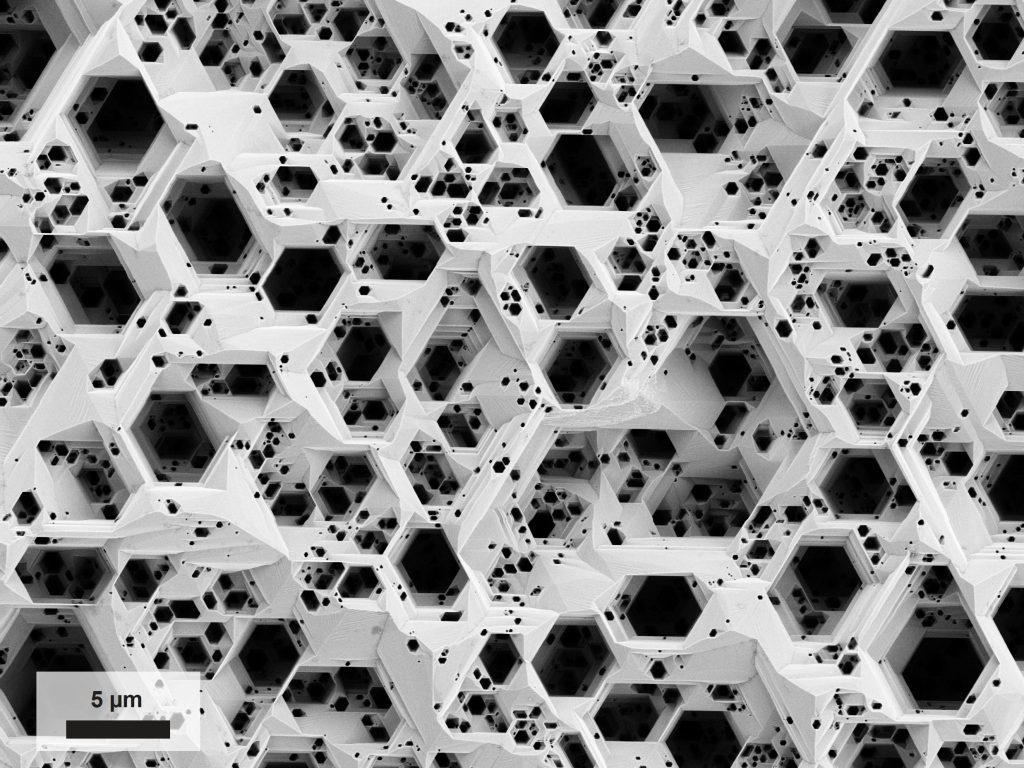
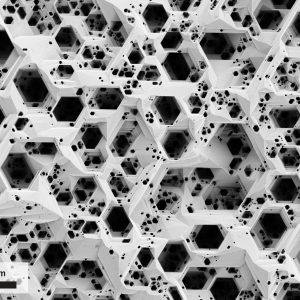
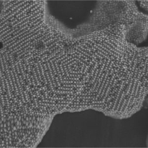
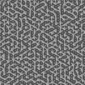
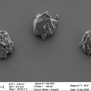
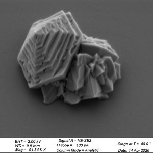
This chaotic “honeycomb” was not sculpted by bees practicing abstract art, but by chemistry. This is a Gallium Nitride wurtzite crystal after aggressive chemical etching, which naturally eats away along the material’s hexagonal planes to create a deep geometric labyrinth. Microscope: SEM Merlin.
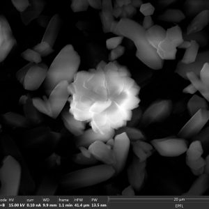
The Crystal Rose, Pratap Narayan Soni (LFIM)
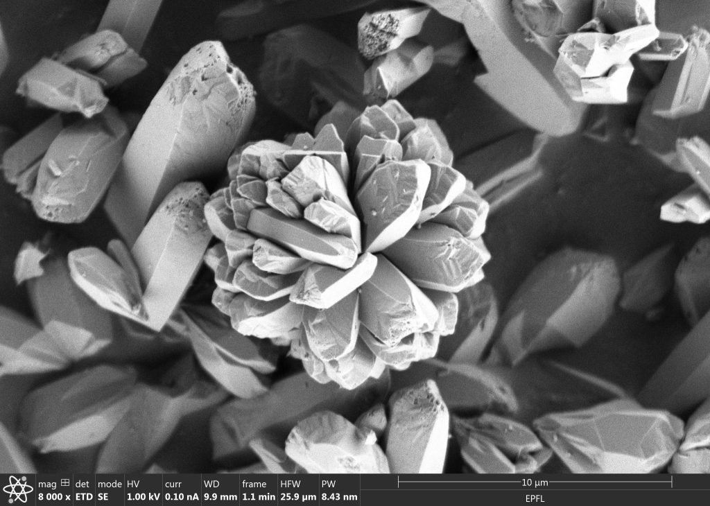
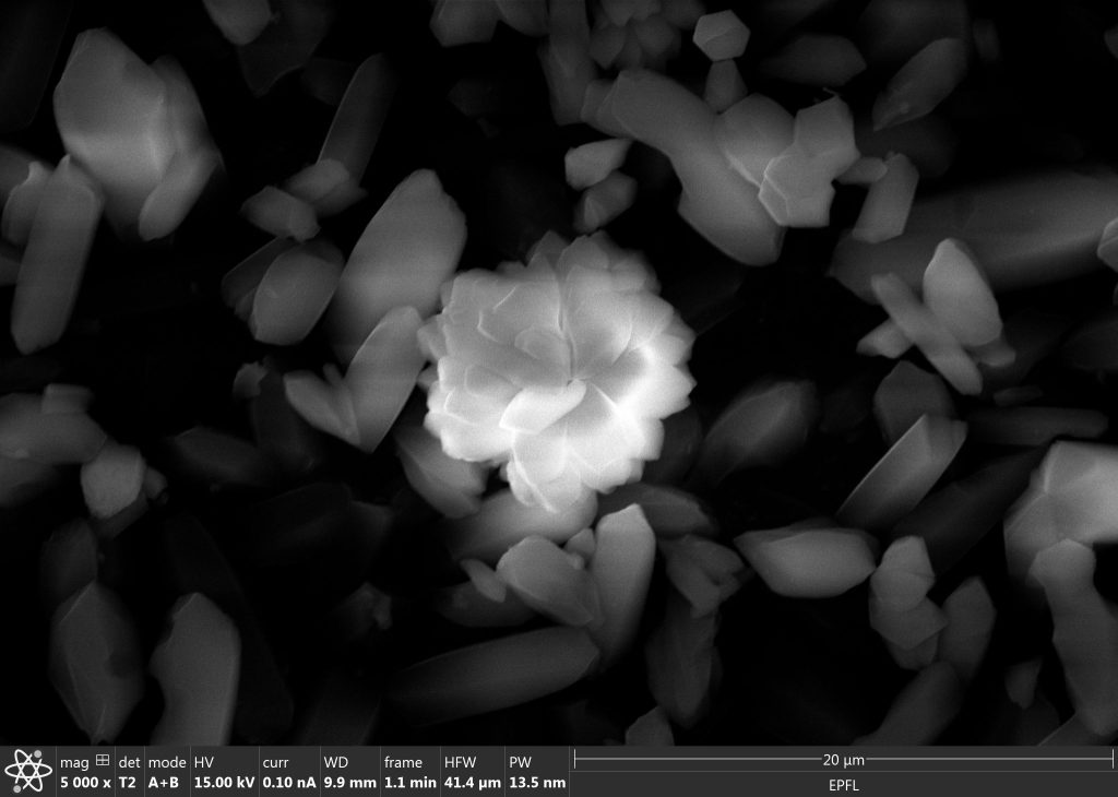
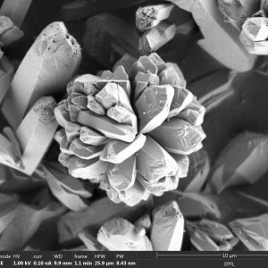
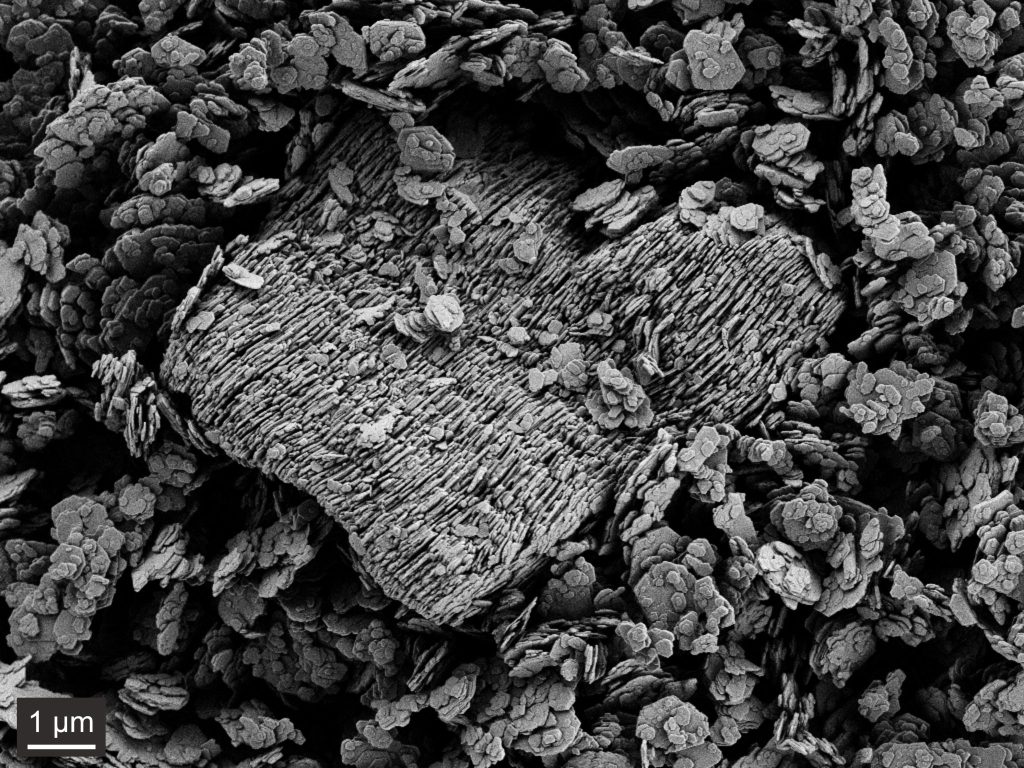
Emerging from a landscape of faceted crystals, this flower-like assembly reveals how microscopic structures can echo forms familiar from nature. Its overlapping crystalline layers resemble the petals of a rose—showing that beauty can flourish even at the micrometre scale. Microscope: SEM Teneo.
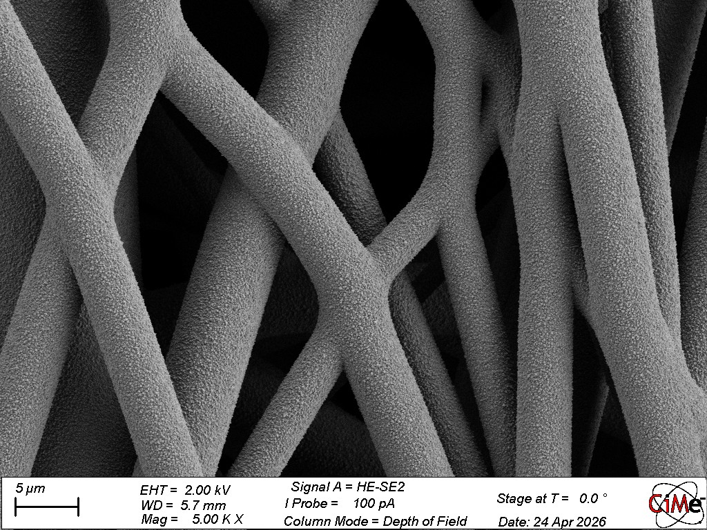
PES-cific Reef, Ceren Kocaman (LAS)
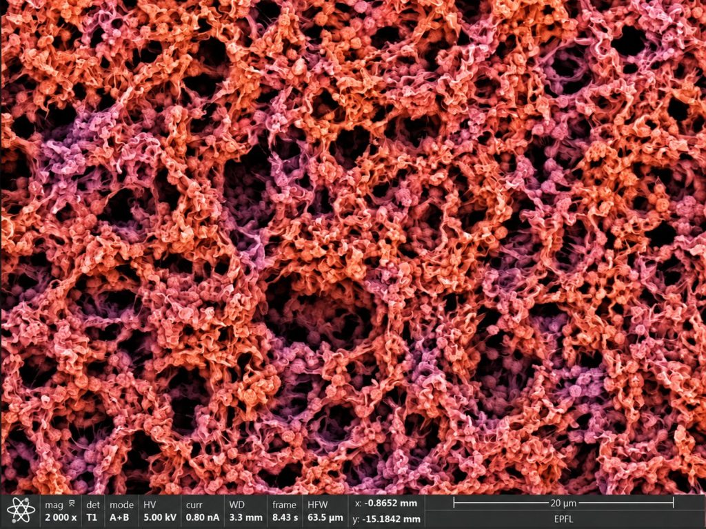
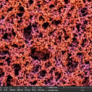
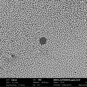
An underwater world at the microscale: this coral-like landscape is actually an interconnected, porous polyethersulfone (PES) network formed by phase inversion. No fish live here; only countless pathways for transport! Microscope: SEM Teneo.
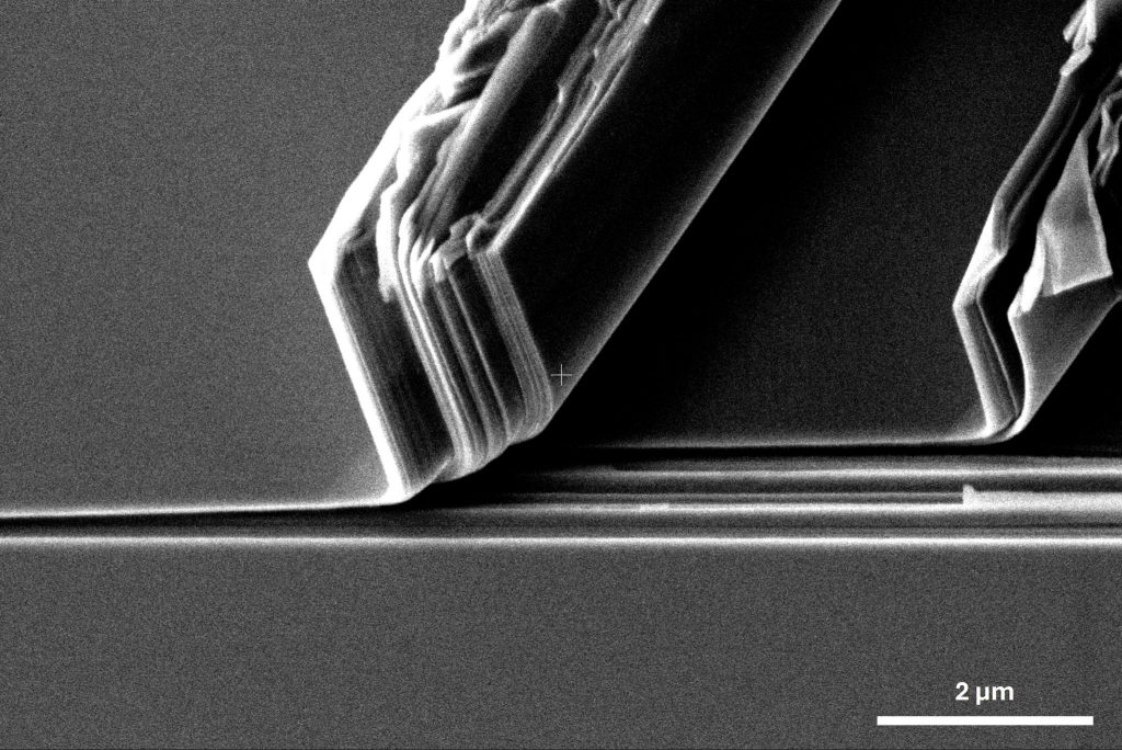
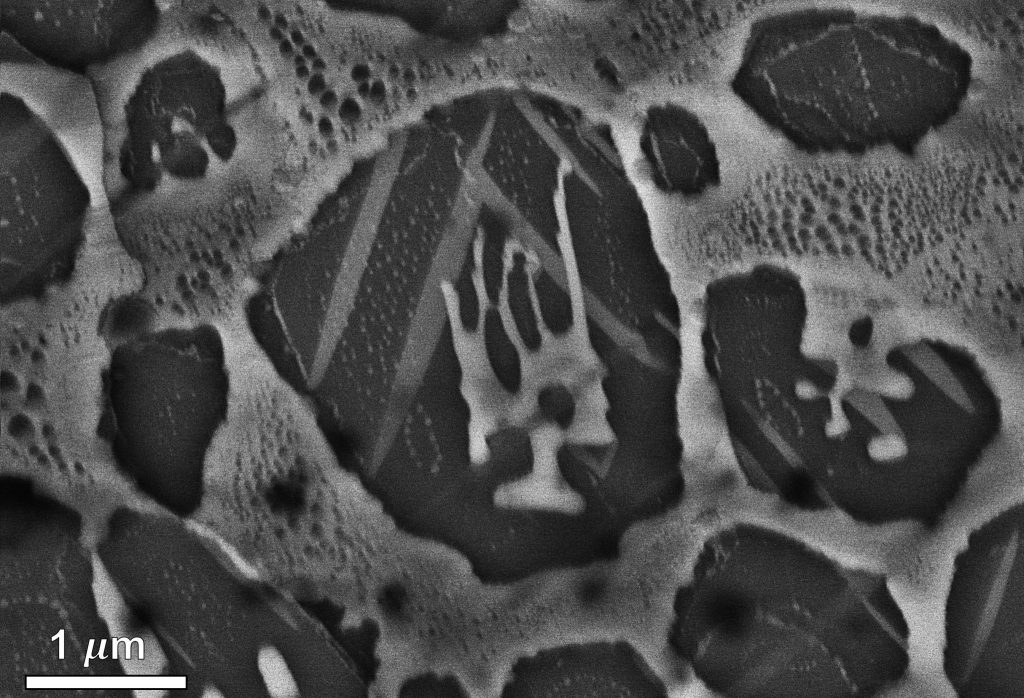
The Shear Signature, Rishi Raj (LPAC)
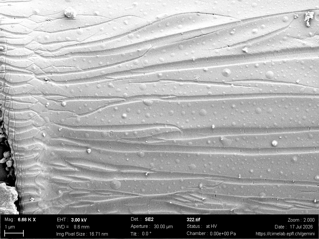
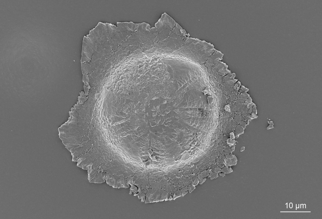
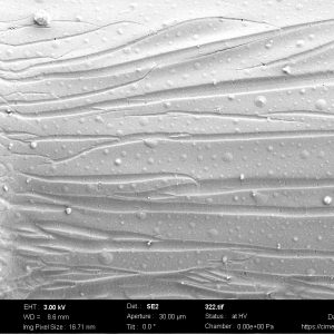
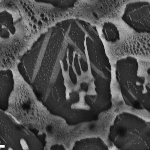
The Micrograph is the edge of a resin microdroplet following an Interfacial Shear Strength (IFSS) test on a single glass fiber. The edge of the microdroplet preserves a remarkable record of the interfacial loading history. The parallel shear lines are the signature residues of stress transfer during single-fiber/microdroplet debonding, illustrating a spatial map of the interface’s response to shear forces and the propagation through the fiber–matrix interface before separation. Microscope: SEM Gemini.
Sunflower, Yan Meng (FIMAP)
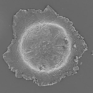
Air bubbles appear on the surface of a polymer thin film, resembling a sunflower or the sun with shining light. Samples come from Dr. Stella Laperrousaz. Microscope: SEM Gemini.
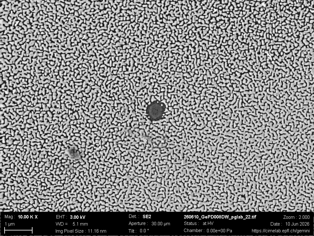
The bark side of dewetting, Laurène Tribolet (FIMAP)
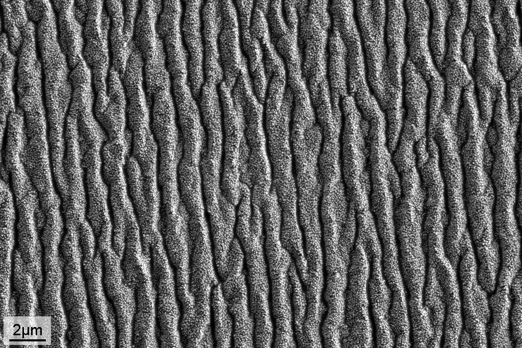
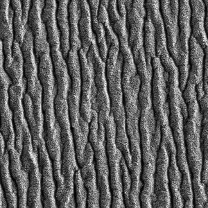
A chalcogenide glass film dewetted on a PDMS substrate whose surface crackled during heating, creating a bark-like texture. Microscope: SEM Gemini.
Germanium in blossom, Ludovica Lunghi (LMSC)
A nanoscale landscape of germanium nanowires, selectively grown and branching across the silicon surface. What appears as a delicate blossom is the result of controlled epitaxial growth, where each structure emerges from the interplay between material, geometry, and growth conditions. Microscope: SEM Gemini.
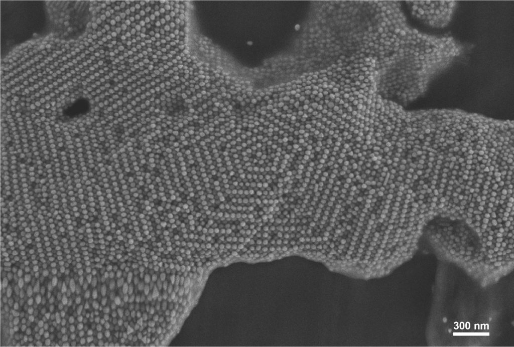
Cuboctahedral gold nanoparticles self-assembled into expanding pentagonal motifs. Microscope: SEM Merlin.
Bloom in the Microcosm, Pratap Narayan Soni (LFIM)
Emerging from a landscape of faceted crystals, this flower-like assembly reveals how microscopic structures can echo forms familiar from nature. Its overlapping crystalline layers resemble the petals of a rose—showing that beauty can flourish even at the micrometre scale. Microscope: SEM Teneo.
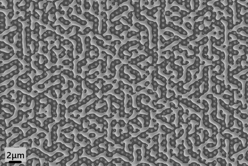
The Chaotic Beehive, Stefano Marinoni (LMSC)
This chaotic “honeycomb” was not sculpted by bees practicing abstract art, but by chemistry. This is a Gallium Nitride wurtzite crystal after aggressive chemical etching, which naturally eats away along the material’s hexagonal planes to create a deep geometric labyrinth. Microscope: SEM Merlin.The Crystal Rose, Pratap Narayan Soni (LFIM)
Emerging from a landscape of faceted crystals, this flower-like assembly reveals how microscopic structures can echo forms familiar from nature. Its overlapping crystalline layers resemble the petals of a rose—showing that beauty can flourish even at the micrometre scale. Microscope: SEM Teneo.PES-cific Reef, Ceren Kocaman (LAS)
An underwater world at the microscale: this coral-like landscape is actually an interconnected, porous polyethersulfone (PES) network formed by phase inversion. No fish live here; only countless pathways for transport! Microscope: SEM Teneo.The Shear Signature, Rishi Raj (LPAC)
The Micrograph is the edge of a resin microdroplet following an Interfacial Shear Strength (IFSS) test on a single glass fiber. The edge of the microdroplet preserves a remarkable record of the interfacial loading history. The parallel shear lines are the signature residues of stress transfer during single-fiber/microdroplet debonding, illustrating a spatial map of the interface’s response to shear forces and the propagation through the fiber–matrix interface before separation. Microscope: SEM Gemini.Sunflower, Yan Meng (FIMAP)
Air bubbles appear on the surface of a polymer thin film, resembling a sunflower or the sun with shining light. Samples come from Dr. Stella Laperrousaz. Microscope: SEM Gemini.The bark side of dewetting, Laurène Tribolet (FIMAP)
A chalcogenide glass film dewetted on a PDMS substrate whose surface crackled during heating, creating a bark-like texture. Microscope: SEM Gemini.Germanium in blossom, Ludovica Lunghi (LMSC)
A nanoscale landscape of germanium nanowires, selectively grown and branching across the silicon surface. What appears as a delicate blossom is the result of controlled epitaxial growth, where each structure emerges from the interplay between material, geometry, and growth conditions. Microscope: SEM Gemini.Pentagonal swirl, Priscila Vensaus & Giulia Dall’Aglio (LNET)
Cuboctahedral gold nanoparticles self-assembled into expanding pentagonal motifs. Microscope: SEM Merlin.Bloom in the Microcosm, Pratap Narayan Soni (LFIM)
Emerging from a landscape of faceted crystals, this flower-like assembly reveals how microscopic structures can echo forms familiar from nature. Its overlapping crystalline layers resemble the petals of a rose—showing that beauty can flourish even at the micrometre scale. Microscope: SEM Teneo.
May/June 2026
“The great book, always open and which we should make an effort to read, is that of Nature” (Antoni Gaudí), Dmitrii Usanov (LQM)
When subjected to mechanical stress, single crystals of layered EuCo2P2 above the brittle-ductile transition point typically bend rather than cleave, revealing sheet-like structures resembling pages. Microscope: FIB/SEM Helios.
The Glass Forest, Nils Golay (LIMNO)
FTO chemical vapor deposition on melted quartz fibers. Microscope: SEM Merlin.
Selenium Maze, Laurène Tribolet & Hugo Hebinger (FIMAP)
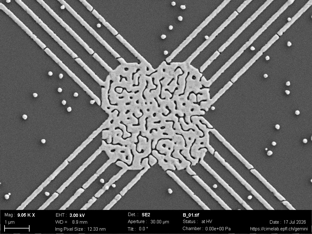
Dewetting of a thin selenium film on a kagome-arranged nanohole array. Microscope: SEM Gemini.
Portrait of the Crowned Platinum Queen in an Intermetallic Kingdom, Mohammadreza Zamani (LMTM)
In this as-cast Pt alloy, no polishing or special preparation was needed for the microstructure to reveal its hidden royalty. The Pt-rich region appears like a crowned Queen’s portrait, embedded in an intermetallic matrix that forms her kingdom. A royal figure, accidentally cast in platinum. Microscope: SEM Gemini.
On-chip Maze Runner, Salvatore Iovinella (LMSC)
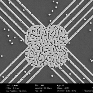
Germanium parasitic epitaxial growth around a contaminant drop on top of a SiO2 mask. The contaminant was probably deposited during a wet process and acted as catalyst during epitaxy. Following the mask roughness, the Ge assumed a labyrinth shape. Microscope: SEM Gemini.
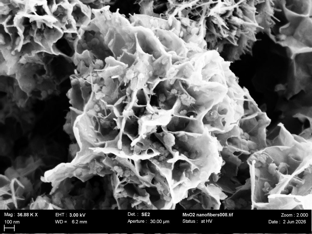
Marigold reef, François Rivat (SMaL)
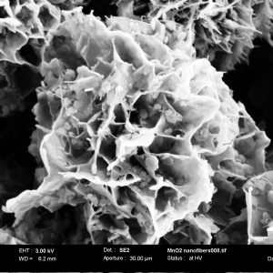
MnO2 urchins obtained after trying to synthesize MnO2 nanofibers. Microscope: SEM Gemini.
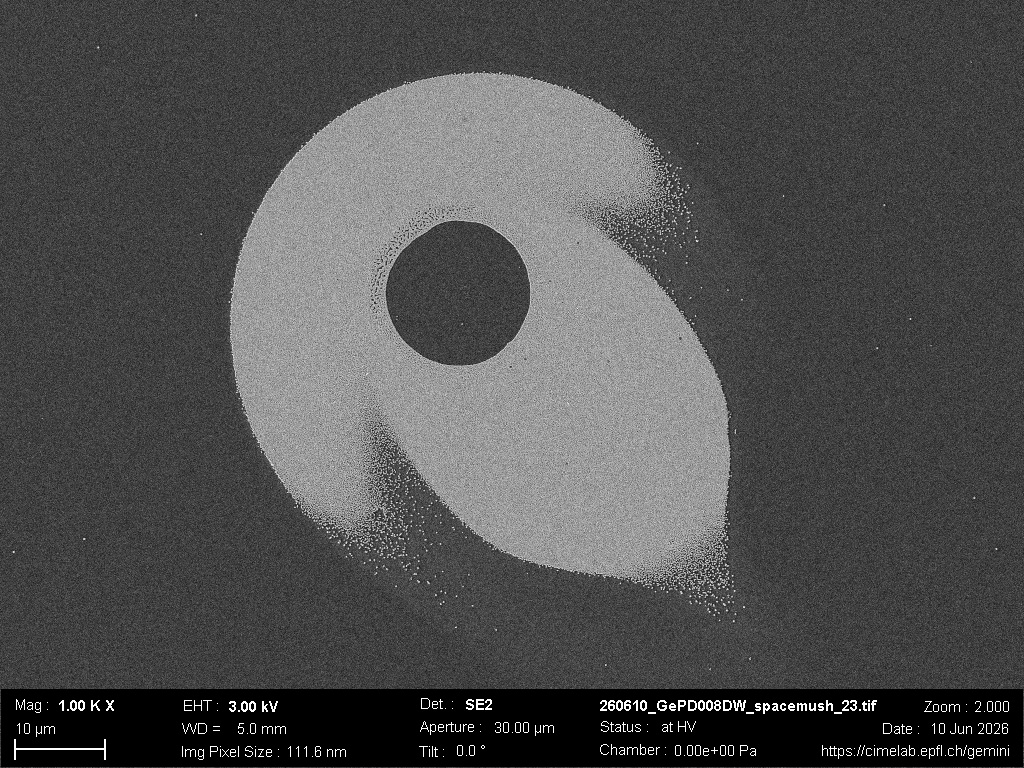
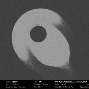
In Orbit Mr.Mushroom, Salvatore Iovinella (LMSC)
Germanium parasitic epitaxial growth around a contaminant drop on top of a SiO2 mask. The contaminant was probably deposited during a fabrication wet process. It acted as a catalyst during epitaxy forming a mushroom shaped shooting star. Microscope: SEM Gemini.
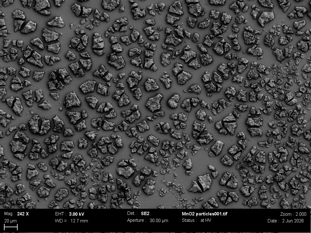
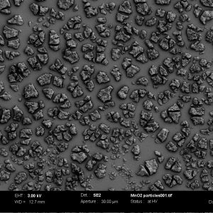
Drylands, François Rivat (SMaL)
MnO2 particles agglomerates that formed upon drying of a dispersion. Microscope: SEM Gemini.
“The great book, always open and which we should make an effort to read, is that of Nature” (Antoni Gaudí), Dmitrii Usanov (LQM)
When subjected to mechanical stress, single crystals of layered EuCo2P2 above the brittle-ductile transition point typically bend rather than cleave, revealing sheet-like structures resembling pages. Microscope: FIB/SEM Helios.The Glass Forest, Nils Golay (LIMNO)
FTO chemical vapor deposition on melted quartz fibers. Microscope: SEM Merlin.Selenium Maze, Laurène Tribolet & Hugo Hebinger (FIMAP)
Dewetting of a thin selenium film on a kagome-arranged nanohole array. Microscope: SEM Gemini.Portrait of the Crowned Platinum Queen in an Intermetallic Kingdom, Mohammadreza Zamani (LMTM)
In this as-cast Pt alloy, no polishing or special preparation was needed for the microstructure to reveal its hidden royalty. The Pt-rich region appears like a crowned Queen’s portrait, embedded in an intermetallic matrix that forms her kingdom. A royal figure, accidentally cast in platinum. Microscope: SEM Gemini.On-chip Maze Runner, Salvatore Iovinella (LMSC)
Germanium parasitic epitaxial growth around a contaminant drop on top of a SiO2 mask. The contaminant was probably deposited during a wet process and acted as catalyst during epitaxy. Following the mask roughness, the Ge assumed a labyrinth shape. Microscope: SEM Gemini.Marigold reef, François Rivat (SMaL)
MnO2 urchins obtained after trying to synthesize MnO2 nanofibers. Microscope: SEM Gemini.In Orbit Mr.Mushroom, Salvatore Iovinella (LMSC)
Germanium parasitic epitaxial growth around a contaminant drop on top of a SiO2 mask. The contaminant was probably deposited during a fabrication wet process. It acted as a catalyst during epitaxy forming a mushroom shaped shooting star. Microscope: SEM Gemini.Drylands, François Rivat (SMaL)
MnO2 particles agglomerates that formed upon drying of a dispersion. Microscope: SEM Gemini.
March/April 2026
Coeur de pierre, Jean-Baptiste Desbrest (LPAC)
Nano platelets of clay, self assembled in the shape of a heart. Microscope: SEM Merlin.
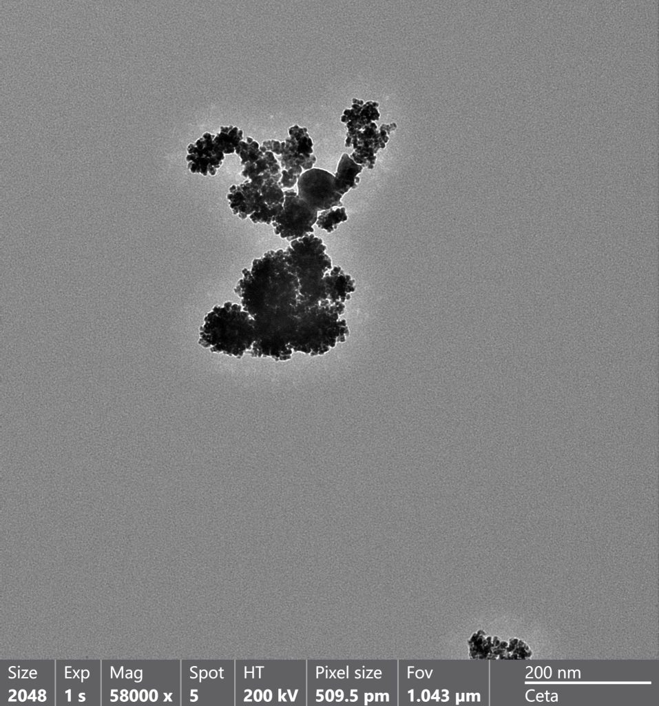
Easter Bunny, Arthur Bouchez and Jiajun Li (LIMNO)
Looks like a cartoon easter bunny jumping out of my material seeking the eggs below. Microscope: TEM Talos.
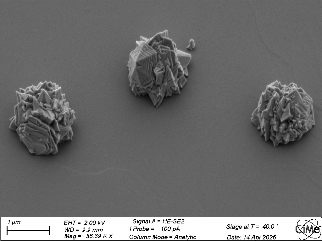
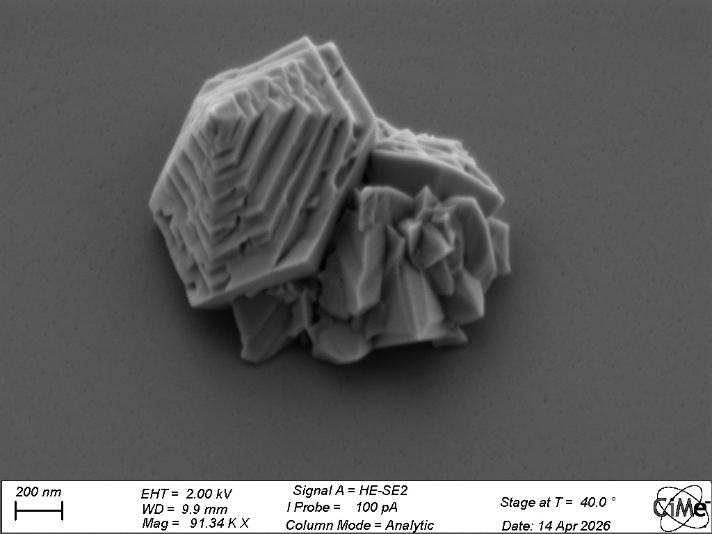
GaN’s and Roses_1, Francesco Marcantoni (POWERLAB)
Under unoptimized MOCVD conditions for Gallium Nitride regrowth, interesting parasitic growth occurs on the SiO2 surface, showing hexagonal structures typical of Wurtzite crystals. Microscope: SEM Merlin.
GaN’s and Roses_2, Francesco Marcantoni (POWERLAB)
Under unoptimized MOCVD conditions for Gallium Nitride regrowth, interesting parasitic growth occurs on the SiO2 surface, showing hexagonal structures typical of Wurtzite crystals. Microscope: SEM Merlin.
The bad bug hiding in the bush, Dea Müller (Bacterial Mechanobiology Laboratory, Persat Lab)
Antibiotic-resistant Acinetobacter baumannii hiding in a flower-like bed of crystallized buffer, exposed by the probing electrons of TEM. Microscope: TEM Bio-Talos.
Coeur de pierre, Jean-Baptiste Desbrest (LPAC)
Nano platelets of clay, self assembled in the shape of a heart. Microscope: SEM Merlin.Easter Bunny, Arthur Bouchez and Jiajun Li (LIMNO)
Looks like a cartoon easter bunny jumping out of my material seeking the eggs below. Microscope: TEM Talos.GaN’s and Roses_1, Francesco Marcantoni (POWERLAB)
Under unoptimized MOCVD conditions for Gallium Nitride regrowth, interesting parasitic growth occurs on the SiO2 surface, showing hexagonal structures typical of Wurtzite crystals. Microscope: SEM Merlin.GaN’s and Roses_2, Francesco Marcantoni (POWERLAB)
Under unoptimized MOCVD conditions for Gallium Nitride regrowth, interesting parasitic growth occurs on the SiO2 surface, showing hexagonal structures typical of Wurtzite crystals. Microscope: SEM Merlin.The bad bug hiding in the bush, Dea Müller (Bacterial Mechanobiology Laboratory, Persat Lab)
Antibiotic-resistant Acinetobacter baumannii hiding in a flower-like bed of crystallized buffer, exposed by the probing electrons of TEM. Microscope: TEM Bio-Talos.
January/February 2026
Trypophobia, Eva Baur (Soft Materials Laboratory)
Structure of porous hydrogels with pores of different length scales. Big pores due to a sacrificial porogen and smaller pores due to the chemical structure of hydrogels. Microscope: SEM Merlin.
HAADF STEM image of 40 nm Cu nanocubes on Cu grid. Microscope: TEM Themis.
A coral in a black ocean, Elisabetta Bonaglia (LPDC)
2% w/w Ni grafted on Al₂O₃, deposited on MgO upon Nickel reduction. The image was taken with the microscope FEI Tecnai Osiris, using HAADF STEM (200 kV, FEI standard single tilt). Microscope: TEM Osiris.
Micro Snow Mountain Range, Yan Meng (FIMAP)
A snow mountain range appears on a curved imprinted polymer thin film. Flat parts are light as the peaks covered by snow while oblique parts are dark like plants. The imprinted structure just offers 3D effect, forming the stereoscopic mountain ridge。Microscope: SEM Gemini.
Lemon race, Laurène Tribolet (FIMAP)
Selenium film dewetting on a substrate patterned with grooves, resulting in lemon-shaped droplets racing in their own track. Microscope: SEM Gemini.
Trypophobia, Eva Baur (Soft Materials Laboratory)
Structure of porous hydrogels with pores of different length scales. Big pores due to a sacrificial porogen and smaller pores due to the chemical structure of hydrogels. Microscope: SEM Merlin.Paragliding Copper Man, Saltanat Toleukhanova (INE)
HAADF STEM image of 40 nm Cu nanocubes on Cu grid. Microscope: TEM Themis.A coral in a black ocean, Elisabetta Bonaglia (LPDC)
2% w/w Ni grafted on Al₂O₃, deposited on MgO upon Nickel reduction. The image was taken with the microscope FEI Tecnai Osiris, using HAADF STEM (200 kV, FEI standard single tilt). Microscope: TEM Osiris.Micro Snow Mountain Range, Yan Meng (FIMAP)
A snow mountain range appears on a curved imprinted polymer thin film. Flat parts are light as the peaks covered by snow while oblique parts are dark like plants. The imprinted structure just offers 3D effect, forming the stereoscopic mountain ridge。Microscope: SEM Gemini.Lemon race, Laurène Tribolet (FIMAP)
Selenium film dewetting on a substrate patterned with grooves, resulting in lemon-shaped droplets racing in their own track. Microscope: SEM Gemini.