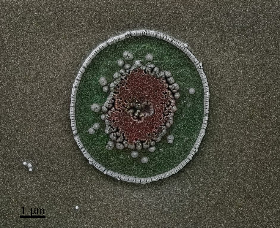
Resembling a predatory piranha bursting from the surface, this SEM image captures the dramatic fracture of a Carbon Nanotube particle. The jagged “teeth” bridging the gaping maw are actually bundles of nanotubes stretched under tension. Microscope: SEM Teneo.
Microscopic Piranha, Yueqing Shen (LAS)
Resembling a predatory piranha bursting from the surface, this SEM image captures the dramatic fracture of a Carbon Nanotube particle. The jagged “teeth” bridging the gaping maw are actually bundles of nanotubes stretched under tension. Microscope: SEM Teneo.
September/October 2025
Edge of the Nano-Forest, Stefano Marinoni (LMSC)
A dense, shadowed forest formed during plasma etching of a Gallium Nitride sample. The tilted SEM view captures its edge, where the trees thin out, revealing the bare substrate beneath. Microscope: SEM Merlin.
Se-pider web, Laurène Tribolet (FIMAP)
A selenium (Se) film caught mid-dewetting over inverted pyramids, weaving a transient web that reflects fluid motion suspended between chaos and order. Microscope: SEM Gemini.
Nanozart and the Symphony in Nano Major, Arthur Bouchez (LIMNO)
A polymer nanoparticle wearing a RuO2 particles wigg ready for a concerto at the TALOS. Microscope: TEM Talos.
Nano-meadow, Stefano Marinoni (LMSC)
This miniature landscape emerged from the controlled chaos of plasma etching. Micromasking transformed a flat gallium nitride surface into a nanoscale meadow — an accidental wilderness of crystalline blades. Microscope: SEM Merlin.
Desert rose, Ariana Serban (LSCI)
Bicarbonate crystals. Microscope: SEM/FIB Helios.
Mickey goes Ga-Ga at the nanoscale, Krishna Kumar (LNCE)
TEM image of Gallium (Ga) nanoparticles mimicking the iconic “Mickey Mouse Silhouette”. Microscope: TEM Spirit.
Pyramids group, Shuqing Song (LAS)
This is a MOF film growing on graphite, with a perfectly orientation. Microscope: SEM Teneo.
Factorial Beehive, Ivan Krsic (POWERlab)
Infinite hexagon inside a hexagon pattern, resembling a beehive, as a result of AlN etching with TMAH. Microscope: SEM Merlin.
Edge of the Nano-Forest, Stefano Marinoni (LMSC)
A dense, shadowed forest formed during plasma etching of a Gallium Nitride sample. The tilted SEM view captures its edge, where the trees thin out, revealing the bare substrate beneath. Microscope: SEM Merlin.Se-pider web, Laurène Tribolet (FIMAP)
A selenium (Se) film caught mid-dewetting over inverted pyramids, weaving a transient web that reflects fluid motion suspended between chaos and order. Microscope: SEM Gemini.Nanozart and the Symphony in Nano Major, Arthur Bouchez (LIMNO)
A polymer nanoparticle wearing a RuO2 particles wigg ready for a concerto at the TALOS. Microscope: TEM Talos.Nano-meadow, Stefano Marinoni (LMSC)
This miniature landscape emerged from the controlled chaos of plasma etching. Micromasking transformed a flat gallium nitride surface into a nanoscale meadow — an accidental wilderness of crystalline blades. Microscope: SEM Merlin.Desert rose, Ariana Serban (LSCI)
Bicarbonate crystals. Microscope: SEM/FIB Helios.Mickey goes Ga-Ga at the nanoscale, Krishna Kumar (LNCE)
TEM image of Gallium (Ga) nanoparticles mimicking the iconic “Mickey Mouse Silhouette”. Microscope: TEM Spirit.Pyramids group, Shuqing Song (LAS)
This is a MOF film growing on graphite, with a perfectly orientation. Microscope: SEM Teneo.Factorial Beehive, Ivan Krsic (POWERlab)
Infinite hexagon inside a hexagon pattern, resembling a beehive, as a result of AlN etching with TMAH. Microscope: SEM Merlin.
July/August 2025
Dinosaur on TEM grid, Richa Rajadhyax (INE)
While searching for my sample, I stumbled upon a little dinosaur instead. Microscope: TEM Talos.
Flowers on desert, Shuqing Song (LAS)
This is a flower shaped ZIF crystal. Microscope: SEM Teneo.
Eye of Sauron, Ariana Serban (LSCI)
Cross-section of nickel fibers covered by Pt/C slurry. Microscope: SEM/FIB Helios.
A boat at the harbour on a sea of hafnia, Niccolò Martinolli (NANOLAB)
The tilted SEM picture displays a portion of gate (left) and contact (right) of a FET fabricated in CMi by Edoardo Tenna, NANOLAB. The TiN gates are patterned first by ion beam etching (IBE), starting from a Si-SiO2-HfO2-TiN stack. The physical Ar etching of the IBE is responsible for the holes and waves left on the hafnia. Some openings are then etched on the hafnia through another photolithography pattern, using buffered HF, followed by the deposition of Ti/Pt contacts by sputtering. The absence of an undercut on the photoresist mask and the sidewall metal deposition typical of the sputtering are responsible for the boat shape. Microscope: SEM Gemini.
Dinosaur on TEM grid, Richa Rajadhyax (INE)
While searching for my sample, I stumbled upon a little dinosaur instead. Microscope: TEM Talos. Flowers on desert, Shuqing Song (LAS)
This is a flower shaped ZIF crystal. Microscope: SEM Teneo.Eye of Sauron, Ariana Serban (LSCI)
Cross-section of nickel fibers covered by Pt/C slurry. Microscope: SEM/FIB Helios.A boat at the harbour on a sea of hafnia, Niccolò Martinolli (NANOLAB)
The tilted SEM picture displays a portion of gate (left) and contact (right) of a FET fabricated in CMi by Edoardo Tenna, NANOLAB. The TiN gates are patterned first by ion beam etching (IBE), starting from a Si-SiO2-HfO2-TiN stack. The physical Ar etching of the IBE is responsible for the holes and waves left on the hafnia. Some openings are then etched on the hafnia through another photolithography pattern, using buffered HF, followed by the deposition of Ti/Pt contacts by sputtering. The absence of an undercut on the photoresist mask and the sidewall metal deposition typical of the sputtering are responsible for the boat shape. Microscope: SEM Gemini.
May/June 2025
Ge snowflake, Ludovica Lunghi (LMSC)
Ge nanowires grown with MOVPE in trenches prepared by e-beam exposure behave as snowflakes do: you can not find two identical ones! Microscope: SEM Gemini.
Walls of pyramid, Shuqing Song (LAS)
The image attached is the surface view of a superlarge grain-MOF membrane.With a SC domain size larger than 5 mircron. Microscope: SEM Teneo.
Golden Domains, Pierpaolo Ranieri (INE)
This TEM image reveals the intricate ferroelectric domain structure of a potassium niobate lamella following a thermal cycle. The pattern is false-colored in golden yellow to highlight the dynamic microstructural beauty of this functional material. Microscope: TEM Talos.
Flash Sutures Ultrafast Molecular Wiring Across a Broken Domain, Ranadip Goswami (LAS)
What once was broken now binds stronger—this SEM image captures the extraordinary act of molecular healing, where ultrafast-growing covalent organic framework (COF) wires thread through a fractured film like nanoscale sutures. The fibrous COF structures emerge from both edges, weaving across the rupture to form a seamless bridge, echoing nature’s own strategies for repair. Microscope: SEM Teneo.
Ge snowflake, Ludovica Lunghi (LMSC)
Ge nanowires grown with MOVPE in trenches prepared by e-beam exposure behave as snowflakes do: you can not find two identical ones! Microscope: SEM Gemini. Walls of pyramid, Shuqing Song (LAS)
The image attached is the surface view of a superlarge grain-MOF membrane.With a SC domain size larger than 5 mircron. Microscope: SEM Teneo.Golden Domains, Pierpaolo Ranieri (INE)
This TEM image reveals the intricate ferroelectric domain structure of a potassium niobate lamella following a thermal cycle. The pattern is false-colored in golden yellow to highlight the dynamic microstructural beauty of this functional material. Microscope: TEM Talos.Flash Sutures Ultrafast Molecular Wiring Across a Broken Domain, Ranadip Goswami (LAS)
What once was broken now binds stronger—this SEM image captures the extraordinary act of molecular healing, where ultrafast-growing covalent organic framework (COF) wires thread through a fractured film like nanoscale sutures. The fibrous COF structures emerge from both edges, weaving across the rupture to form a seamless bridge, echoing nature’s own strategies for repair. Microscope: SEM Teneo.
March/April 2025
Red House on Cracked Green Earth, Qiucheng Xu (LSCI)
False‐colored SEM micrograph of a carbon gas diffusion electrode (GDE) surface, acquired on a field‐emission scanning electron microscope at 10 kV with the sample stage tilted ~25° to accentuate the microporous layer’s topography. The carbon surface is rendered in green and ochre tones, deep crevices appear in dark brown, and an isolated particulate feature has been false-colored bright red to resemble a small house perched above the cracked “landscape.” Microscope: SEM Gemini.
Easter Egg, Pierre-Luc Piveteau (STI-IMX-FIMAP)
Similar to water, which is known to form beads on superhydrophobic surfaces, a quenched chalcogenide glass can form a high contact-angle dropplet on a fluorosilane-treated surface. This “egg” was not brought by the bunny, but was rather caused by the dewetting of the glass-melt layer from the surface. Microscope: SEM Gemini.
Molecular elegance hexagonal DNA arrays in a Moiré performance, Anna Duvakina (LMGN)
Hexagonal DNA 2D arrays fold over the TEM grid, layering into a Moiré pattern — demonstrating how a subtle twist can give rise to a work of art. Microscope: TEM Bio-Talos.
Micro xmas biscuits, Thi Ha My Pham (LMER)
Metal-organic cages. Microscope: SEM Teneo.
Fly muscle, Stéphanie Clerc-Rosset (PTBioEM)
TEM image of a single muscle fibre from a fruit fly. Microscope: TEM Bio-Spirit.
Dinosaur on TEM grid, Richa Rajadhyax (INE)
While trying to locate my sample I stumbled upon a little dino playing hide and seek. Microscope: TEM Talos.
A crystalline flower, Thi Ha My Pham (LMER)
Metal-organic cages forming crystalline flower shape. Microscope: SEM Teneo.
Red House on Cracked Green Earth, Qiucheng Xu (LSCI)
False‐colored SEM micrograph of a carbon gas diffusion electrode (GDE) surface, acquired on a field‐emission scanning electron microscope at 10 kV with the sample stage tilted ~25° to accentuate the microporous layer’s topography. The carbon surface is rendered in green and ochre tones, deep crevices appear in dark brown, and an isolated particulate feature has been false-colored bright red to resemble a small house perched above the cracked “landscape.” Microscope: SEM Gemini.Easter Egg, Pierre-Luc Piveteau (STI-IMX-FIMAP)
Similar to water, which is known to form beads on superhydrophobic surfaces, a quenched chalcogenide glass can form a high contact-angle dropplet on a fluorosilane-treated surface. This “egg” was not brought by the bunny, but was rather caused by the dewetting of the glass-melt layer from the surface. Microscope: SEM Gemini.Molecular elegance hexagonal DNA arrays in a Moiré performance, Anna Duvakina (LMGN)
Hexagonal DNA 2D arrays fold over the TEM grid, layering into a Moiré pattern — demonstrating how a subtle twist can give rise to a work of art. Microscope: TEM Bio-Talos.Micro xmas biscuits, Thi Ha My Pham (LMER)
Metal-organic cages. Microscope: SEM Teneo.Fly muscle, Stéphanie Clerc-Rosset (PTBioEM)
TEM image of a single muscle fibre from a fruit fly. Microscope: TEM Bio-Spirit.Dinosaur on TEM grid, Richa Rajadhyax (INE)
While trying to locate my sample I stumbled upon a little dino playing hide and seek. Microscope: TEM Talos.A crystalline flower, Thi Ha My Pham (LMER)
Metal-organic cages forming crystalline flower shape. Microscope: SEM Teneo.
January/February 2025
Nanoprinted Rolex Learning Center- A Fusion of Art and Nanotechnology_Huixin Guo (LMGN)
This image showcases a nanoprinted miniature of EPFL’s iconic Rolex Learning Center, fabricated at CMI. The structure was created using two-photon lithography (Nanoscribe) to pattern a polymer template, followed by atomic layer deposition (ALD) to coat it with nickel (Ni), replicating the architectural curves with nanoscale precision. This work exemplifies the power of advanced nanofabrication techniques, merging architectural beauty with cutting-edge material science. Microscope: SEM Merlin.
Wooden Sign-board_Archisman Ghosh (LAS)
A beautifully shaped zeolite crystal which resembles a wooden road signboard. Microscope: SEM Teneo.
The golden train has come!_Mohammad Rezaei (POWERlab)
Get shrunk and get in. Except the oversized ones, everyone is welcome. The terrain is nonconductive, but don’t worry, the drive will conduct well the train, even in 75 (45+30) degree slopes … Microscope: SEM Merlin.
Tiny Mineral_Yan Meng (FIMAP)
Small chalcogenide glass fragments suspending on the polymer substrate look like tiny mineral grown in the cave. Microscope: SEM Gemini.
Spider nest_Sönke Menke (LMIS1)
While investigating my samples, I stumbled upon this area that looks like some micro-spiders have build their nest and deposited some eggs here. Microscope: SEM Gemini.
The Great Coral Reef of Lausanne_Sönke Menke (LMIS1)
While thinking about summer vacation, I stumbled upon this section of my sample, that reminded me of a coral reef. Maybe this will help fight the winter blues until we see more sun! Microscope: SEM Gemini.
Glowstone cave_Sönke Menke (LMIS1)
Sometimes, while exploring the unknown, one might stumble upon caves filles with precious glowstones. Be quick to pick them up for your future journey. Microscope: SEM Gemini.
Space invaders- VO2 edition_Niccolò Martinolli (NANOLAB)
FIB cross-section of a 160 nm VO2 nanowire and 60 nm VO2 bottom layer on top of a Si chip with 200 nm SiO2 and covered with 20 nm Cr and 150 nm Au contact. The nanowire has been obtained by ion beam etching (IBE) with Ar, using 900 nm HSQ (e-beam lithography resist) as a mask. The combination of the high aspect ratio, the typical IBE redeposition, and the -10° angle and rotation during the etching has produced the antenna-like VO2 structures. The chip was fabricated by Vanessa Conti and Andrea Iaconeta in CMi, the FIB lamella and TEM analysis have been done by me. The space invader aliens have been added in post-production. Microscope: TEM Talos.
Sea-lenium Waves_Laurène Tribolet (FIMAP)
A defect in a cured transparent resist created wrinkles, forming wave-like structures. During the dewetting of a thin selenium film, droplets formed and settled on the surface, appearing to float on the waves. Microscope: SEM Gemini.
Coffins lying around nr.1_Archisman Ghosh (LAS)
An SEM image of coffin-shaped zeolite crystals. Microscope: SEM Teneo.
Coffins lying around nr.2_Archisman Ghosh (LAS)
Some zeolite crystals deposited on graphite closely resemble a bunch of coffins. Microscope: SEM Teneo.
Breathing Heart- The Porous Bloom_Ranadip Goswami (LAS)
A heart-shaped structure intricately designed from covalent organic framework nanotubes, resembling a blooming flower—an elegant fusion of porosity and precision that breathes life into material science. Microscope: SEM Teneo.
The Triple-Walled InP Fortress, Riccardo Brondolin (LMSC)
nP grown my MBE turns contamination into fascinating art – shaping intricate, fortress-like layers that echo medieval defenses. Microscope: SEM Merlin.
Nanoprinted Rolex Learning Center- A Fusion of Art and Nanotechnology_Huixin Guo (LMGN)
This image showcases a nanoprinted miniature of EPFL’s iconic Rolex Learning Center, fabricated at CMI. The structure was created using two-photon lithography (Nanoscribe) to pattern a polymer template, followed by atomic layer deposition (ALD) to coat it with nickel (Ni), replicating the architectural curves with nanoscale precision. This work exemplifies the power of advanced nanofabrication techniques, merging architectural beauty with cutting-edge material science. Microscope: SEM Merlin.Wooden Sign-board_Archisman Ghosh (LAS)
A beautifully shaped zeolite crystal which resembles a wooden road signboard. Microscope: SEM Teneo.The golden train has come!_Mohammad Rezaei (POWERlab)
Get shrunk and get in. Except the oversized ones, everyone is welcome. The terrain is nonconductive, but don’t worry, the drive will conduct well the train, even in 75 (45+30) degree slopes … Microscope: SEM Merlin.Tiny Mineral_Yan Meng (FIMAP)
Small chalcogenide glass fragments suspending on the polymer substrate look like tiny mineral grown in the cave. Microscope: SEM Gemini.Spider nest_Sönke Menke (LMIS1)
While investigating my samples, I stumbled upon this area that looks like some micro-spiders have build their nest and deposited some eggs here. Microscope: SEM Gemini.The Great Coral Reef of Lausanne_Sönke Menke (LMIS1)
While thinking about summer vacation, I stumbled upon this section of my sample, that reminded me of a coral reef. Maybe this will help fight the winter blues until we see more sun! Microscope: SEM Gemini.Glowstone cave_Sönke Menke (LMIS1)
Sometimes, while exploring the unknown, one might stumble upon caves filles with precious glowstones. Be quick to pick them up for your future journey. Microscope: SEM Gemini.Space invaders- VO2 edition_Niccolò Martinolli (NANOLAB)
FIB cross-section of a 160 nm VO2 nanowire and 60 nm VO2 bottom layer on top of a Si chip with 200 nm SiO2 and covered with 20 nm Cr and 150 nm Au contact. The nanowire has been obtained by ion beam etching (IBE) with Ar, using 900 nm HSQ (e-beam lithography resist) as a mask. The combination of the high aspect ratio, the typical IBE redeposition, and the -10° angle and rotation during the etching has produced the antenna-like VO2 structures. The chip was fabricated by Vanessa Conti and Andrea Iaconeta in CMi, the FIB lamella and TEM analysis have been done by me. The space invader aliens have been added in post-production. Microscope: TEM Talos.Sea-lenium Waves_Laurène Tribolet (FIMAP)
A defect in a cured transparent resist created wrinkles, forming wave-like structures. During the dewetting of a thin selenium film, droplets formed and settled on the surface, appearing to float on the waves. Microscope: SEM Gemini.Coffins lying around nr.1_Archisman Ghosh (LAS)
An SEM image of coffin-shaped zeolite crystals. Microscope: SEM Teneo.Coffins lying around nr.2_Archisman Ghosh (LAS)
Some zeolite crystals deposited on graphite closely resemble a bunch of coffins. Microscope: SEM Teneo.Breathing Heart- The Porous Bloom_Ranadip Goswami (LAS)
A heart-shaped structure intricately designed from covalent organic framework nanotubes, resembling a blooming flower—an elegant fusion of porosity and precision that breathes life into material science. Microscope: SEM Teneo.The Triple-Walled InP Fortress, Riccardo Brondolin (LMSC)
nP grown my MBE turns contamination into fascinating art – shaping intricate, fortress-like layers that echo medieval defenses. Microscope: SEM Merlin.