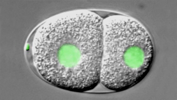
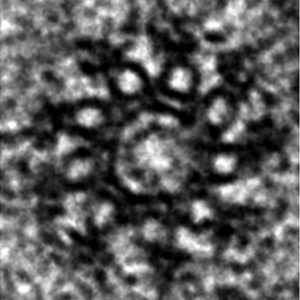
Deciphering and engineering the assembly of cellular organelles is a frontier pursuit in biology. Centrioles are small cylindrical organelles essential for the formation of cilia, flagella and centrosomes, and characterized by a remarkable nine-fold radial symmetric arrangement of microtubule triplets. How centrioles assemble, usually once and only once per cell cycle, has been a long-standing question in cell and developmental biology. Over a decade ago, forward genetic and functional genomics screens identified a small set of proteins essential for the onset of centriole assembly across evolution, whose mechanism of action is being elucidated. In most systems, centrioles assemble around a 9-fold symmetrical cartwheel structure next to each resident centriole. In human cells, the cartwheel emerges from a torus encircling the proximal end of each resident centriole, to which the proteins PLK4, STIL and HsSAS-6 are recruited.
A working model suggests that PLK4 and associated STIL focus to a single location on this torus, which might enable HsSAS-6 concentration and cartwheel emergence. We proposed a structural model in which the association of 9 homodimers of SAS-6 proteins to form a central ring from which radiate nine spokes is at the root of the near-universal 9-fold symmetry of centrioles. We obtained evidence supporting this model using high-speed Atomic Force Microscopy (AFM), in collaboration with the laboratory of Georg Fantner (EPFL). Furthermore, we developed a cell-free assay that enabled us to uncover that SAS-6 proteins possess an autonomous ability to form cartwheel-like structures. In related work, we are interested in understanding how centrioles are eliminated in some circumstances, including during oogenesis.
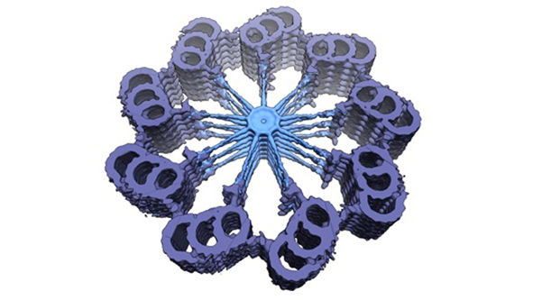
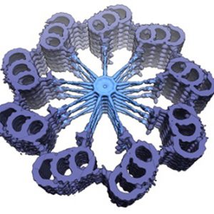
Watch movie of slightly tilted cross-sectional 3D map of the proximal region of the centriole in Trichonympha ssp., illustrating the striking 9-fold radial symmetry of the organelle. The cartwheel structure with stacks of SAS-6-bearing rings is visible in light blue, the more peripheral pinhead element in dark blue. The most peripheral microtubule triplets are shown in purple and the A-C linker connecting them in green. The cross-sectional diameter is ~250 nm.