Our group, in collaboration with the EPFL School of Engineering, Institute of Electrical and Micro Engineering and the EPFL School of Life Science, successfully demonstrates growth of a network of living and functional primary mouse hippocampal neurons on a nanostructured single crystal diamond surface. Our results suggest that neuron growth can be tailored on diamond nanopillars to realize a nanophotonic quantum sensing platform for wide-field and label-free neuronal activity recording with sub-cellular resolution.
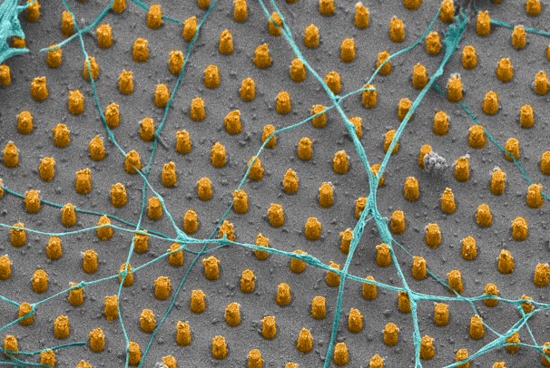
The sensing mechanism is based on Nitrogen-Vacancy (NV) centers in diamond consisting of a nearest-neighbor pair of a nitrogen atom (substituting for a carbon atom) and a lattice vacancy. These intrinsic defect centers can be used for remote sensing of magnetic and electric field at sub-micrometer resolution, thanks to optical polarization and readout of their spin states by optically detected magnetic resonance. In order to increase the photoluminescence collection efficiency and to assure the best contact between the cells and the diamond, we demonstrate a highly controllable and repeatable nanofabrication process for producing high aspect ratio, large scale nanopillar arrays made of single crystal diamond.
Taking advantage of the NVs naturally present in our sample, we verify enhanced photoluminescence from pillar locations while spin resonance spectra remain unaffected by the nanostructuring. In addition, we thoroughly characterize the neurons grown on the nanostructured diamond surfaces. We demonstrate with electrophysiology measurements that the neurons are alive and functional. Using scanning electron microscopy, we investigate the relative position between the cells and the pillars at the nanometer scale. Moreover, we establish how neurons grow depending on varying geometrical parameters of the array.
Our work demonstrates that the envisioned sensing platform is compatible with functional neurons, which is a milestone towards an NV-based sensing platform for living neurons. Once realized, this sensing platform would offer the possibility to monitor neuronal activity with simultaneously high spatial and temporal resolution, which is crucial to advance understanding of the development and functioning of our brain, and to gain further insights in the origin of brain disorders.

Reference: “Neuronal growth on high‑aspect‑ratio diamond nanopillar arrays for biosensing applications”, Elena Losero, Somanath Jagannath, Maurizio Pezzoli, Valentin Goblot, Hossein Babashah, Hilal A. Lashuel, Christophe Galland & Niels Quack, Scientific Reports, 13, 5909 (2023); https://www.nature.com/articles/s41598-023-32235-x