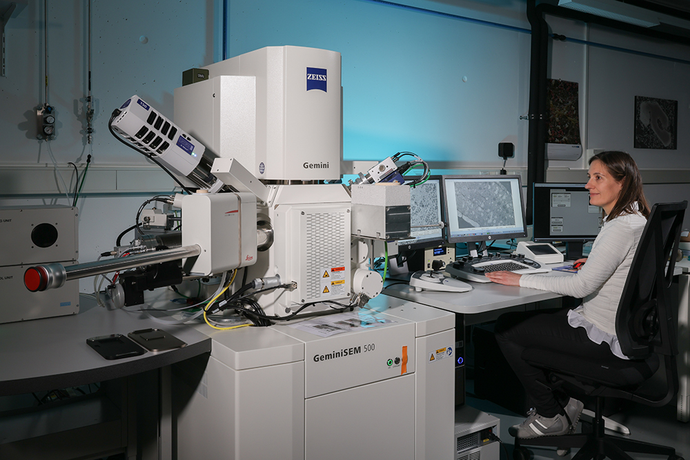
A new state-of-the-art cryo scanning electron microscope (cryo-SEM), a Gemini 500, was acquired between the Faculty of Architecture, Civil- and Environmental Engineering at EPFL and the Faculty of Geology and Environmental Sciences at the University of Lausanne in 2017. The Gemini 500 SEM instrument is now in service within the Center for Advanced Surface Analysis (CASA), established between EPFL and the University of Lausanne since 2013, operated by laboratory manager Dr. Cristina Martin-Olmos reporting to Prof. Anders Meibom.
The Gemini 500 SEM instrument can analyze a wide range of samples, including biological tissue, geological specimens, fossils, soft materials, polymers, nanoparticles, electronics, and microstructures of e.g. building materials.
SEM is a surface technique, but with careful sample preparation such as fracture- and polishing techniques, ultramicrotomy (coupled with Scanning Transmission Electron Microscopy, STEM) or array tomography, we can also access the volume of small samples. Furthermore, material contrast via back scattered electrons (BSE); elemental analysis with energy dispersive x-ray spectroscopy (EDX), and charge contrast imaging can allow us to distinguish different materials within a given sample specimen.
Technical overview
SEM:
- Electron source: Schottky FEG, 3-20 nAmp.
- Accelerating voltage: 20 V – 30 kV.
- Resolution: 0.9 nm at 1 kV with Tandem Decel, 1.2 nm at 500 V without deceleration, 0.6 nm at 15 kV.
- Electron detectors: In-lens SE and EsB, VP-SE, VP BSE, Everhart-Thornley SE, 7 panel STEM.
- Analysis: Oxford AZTec Advanced Microanalysis system with X-MAX 150mm2 detector with 127 eV (@ Mn Ka).
- Techniques: SEM (SE & BSE), EsB, charge contrast imaging, electron channeling, EsB, EDX, Cryo-SEM, VP-SEM (N2 and H2O), STEM: BF, DF, ADF & HAADF.
Attachments:
- Cryo stage and cryo transfer system, Leica VCT500.
- Low kV Argon ion gun.
- NanoVP.
- Plasma cleaner.
- Peltier cooling stage.
- 80 mm Air-lock.
- 2 CCD cameras.
Preparation:
- Ultramicrotomy and cryo-ultramicrotomy.
- Specialized diamond knives for array tomography.
- Axio Imager M2. Stage and software for correlative microscopy.
- VCT500 cryo-transfer system, also connected to the CryoNanoSIMS.
- E-beam coating, room temperature and cryo-conditions with Pt and C.
- Freeze fracture.
- Freeze drying.
- Glow discharge.
Description:
The Gemini500 SEM was the first of its kind in Switzerland. The instrument is especially well-suited for low-kV SEM. The efficiency of the in-lens detectors creates crisp images with low noise, even at very low kV. The microscope has a resolution of 1.2 nm at 500 V without beam deceleration, and 0.9 nm at 1 kV with Tandem Decel beam deceleration. This minimizes electron beam damage to sensitive specimens and facilitates working with uncoated samples. The microscope is equipped with smartStitch large area imaging and stitching software.
The energy selective backscatter detector (EsB) is especially useful for material contrast at low kV and allows for the repelling of secondary electrons and imaging with low-loss back scattered electrons. The Gemini500 features simultaneous imaging with the in-lens secondary and in-lens backscatter detector.
Furthermore, thanks to a NanoVP mode, it is possible to use the in-lens detectors in variable pressure (VP) mode, i.e. acquire images in the presence of nitrogen gas at up to 150 Pa with high resolution (e.g. 1.8 nm at 3kV, 30 Pa). This permits imaging of uncoated samples without charging.
The microscope also has VP SE and VP BSE detectors for pressures of up to 500 Pa and a classic Everhart-Thornley detector for high vacuum SE. For sections on grids the microscope is fitted with a multi-panel a-STEM4 detector with parallel read-out of 4 virtual STEM modes. For elemental analysis, there is an Oxford EDX with a 150 mm2 X-MAX detector
The microscope has a cryo-stage, a cryo shield, and a vacuum cryo-transfer system. This permits contamination free transfer of frozen samples to the chamber and imaging of samples at temperatures down to -180 °C.
Unique features:
Our Gemini500 is equipped with water vapor for the VP mode, thus it features both nitrogen and water. Combining a partial water vapor pressure of 500 Pa with a Peltier cooling stage (-30 °C to 45 °C) that keeps the sample temperature just above 0 °C permits to reach a relative humidity of ~80 %. This means that we can image hydrated samples for extended time with only slow dehydration.
Furthermore, as the only cryo-SEM in the world, this Gemini500 is equipped with a low kV ion Argon gun attached directly on the chamber, allowing to clean the surface of cryo-samples directly inside the microscope chamber.
Preparation:
For the preparation of frozen samples our lab is equipped with a cryo-ultramicrotome, Leica FC7/UC7 with micromanipulators for CEMOVIS, a mounting station for handling samples under liquid nitrogen, Leica VMC, and a cryo-vacuum chamber, Leica ACE600, for performing freeze-fracture, freeze drying and e-beam coating (Pt and C). The ACE600 is also operational in room temperature and features a glow discharge unit.
Furthermore, we have two room temperature ultramicrotomes and a Zeiss Axio Imager M2, which works in transmission and reflection mode, has circular DIC and a holder and software for correlative microscopy of sections on Si wafers.