Spatial and Temporal Focusing
Two-photon fluorescent excitation (TPFE) imaging through optical fibers has traditionally been based on double clad fibers or fiber bundles. In the double clad fiber systems, the single mode core is used to deliver illumination. A lens placed at the distal tip of the fiber focuses the light and a scanning mechanism is required at the distal tip to move the focused spot over the field of view. The two-photon fluorescence is collected through the cladding. In the case of the fiber bundle, the cores introduce pixilation to the image and limit the resolution of the imaging device due to the fact that each of the individual cores of the bundle acts as an individual “pixel” that relays the image through the fiber.
For the same fiber diameter, the information capacity of multimode optical fibers, given by the large number of supported propagation modes, is larger than that of fiber bundles which potentially allows the transmission of higher resolution images through ultra-thin endoscopic probes.
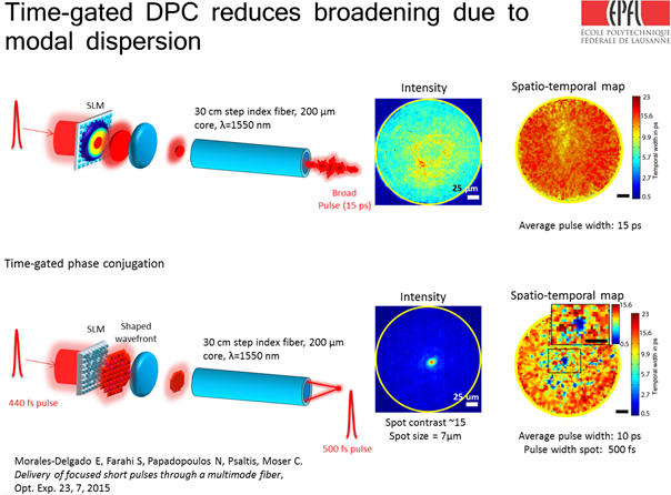
Transmission of ultrashort pulses through multimode fibers generates a scrambled intensity profile and a broadened temporal pulse width due to modal dispersion.
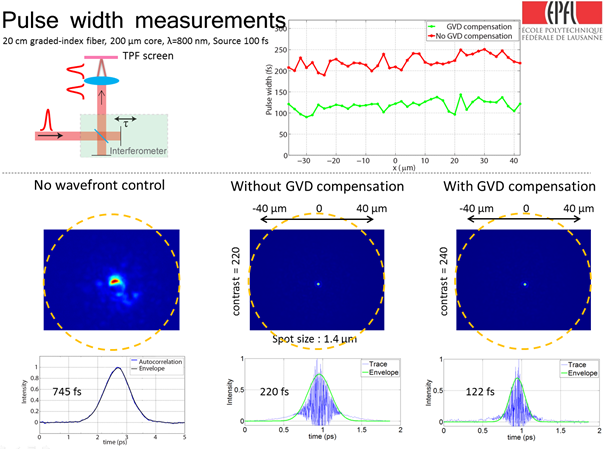
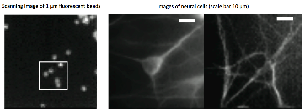
In this investigation we have developed a method to deliver and scan spatially focused femtosecond pulses through multimode optical fibers. We minimize pulse broadening due to modal dispersion by a time-gated mode-selective phase conjugation process, in which only a subset of modes of similar group velocities is counter-propagated through the fiber. Excitation of modes with the same speed reduces broadening due to modal dispersion. Additionally, group velocity dispersion (GVD) is reduced by chirping the pulse. Hence, our method allows the transmission and scanning of near-diffraction-limited focused optical pulses of 120 fs pulse width through a 20 cm length multimode optical fiber. Scanning of the focused pulse and collection through the same fiber of the TPFE signal allows the acquisition of two-photon images of the sample placed at the distal side.
Publications: A. Flusberg, E. D. Cocker, W. Piyawattanametha, J. C. Jung, E. L. M. Cheung, and M. J. Schnitzer, “Fiber-optic fluorescence imaging,” Nature Methods 2, 941-950 (2005).
Bianchi, S. & Di Leonardo, R. A multimode fiber probe for holographic micromanipulation and microscopy. Lab Chip 12, 635–639 (2012).
Čižmár, T. & Dholakia, K. Exploiting multimode waveguides for pure fibre-based imaging. Nat. Commun. 3, 1027 (2012).
N. Papadopoulos, S. Farahi, C. Moser, and D. Psaltis, “High-resolution, lensless endoscope based on digital scanning through a multimode optical fiber,” Biomedical Opt. Express 4, 260 (2013).
Andresen, E. R., Bouwmans, G., Monneret, S. & Rigneault, H. Two-photon lensless endoscope. Optics Express 21, 20713–20721 (2013)
Morales-Delgado, S. Farahi, I. Papadopoulos, D. Psaltis, and C. Moser, “Delivery of focused short pulses through a multimode fiber,” Opt. Express 23, 9109-9120 (2015).
Contact: Edgar Emilio Morales Delgado.
Multimode Fiber Based Endoscopy
Imaging hidden and inaccessible structures has become possible by modern endoscopy with applications in medical diagnosis and treatment as well as in manufacturing where the visualization of hidden structures is important to increase quality of manufactured goods. Today there are two main approaches to build a miniaturized endoscope. The first is the miniaturization of optical elements (micro GRIN lenses) and the second is a fiber-based approach for flexible endoscopes. Both approaches suffer from limitations as the overall size of the device or the low resolution.
In this project, we work on developing an endoscope using a single multimode fiber (MMF) for both light delivery and collection. By taking advantage of the degrees of freedom provided by the multimode fiber and given by the number of its supported modes, we can use a single multimode fiber as a lens-less endoscope with a sub-micrometer resolution.
Thanks to digital phase conjugation we can compensate for modal scrambling inherent to multimode fibers and responsible for image distortion. Indeed, light focused into a MMF is coupled in different modes, which propagate with different propagation constants and interact by coupling energy from one mode into another. This modal scrambling leads to the complete distortion of an input image at the output of the fiber. Even though the image at the output of the MMF is completely distorted, the information of the original image remains and can be recovered.
Once the fiber is calibrated, we can focus on a single spot without the use of a lens and scan the spot by pure digital means by controlling the wave-front injected in the multimode fiber. We acquire images by raster scanning.
Publications:
Papadopoulos I., Farahi S., Moser C., Psaltis D., High-resolution, lensless endoscope based on digital scanning through a multimode optical fiber, Biomed. Opt. Express, Vol. 4, Issue 2, pp. 260-270, 2013.
Papadopoulos I., Farahi, S., Moser C., Psaltis D., Focusing and scanning light through a multimode optical fiber using digital phase conjugation, Optics Express, Vol. 20, Issue 10, pp. 10583-10590, 2012.
Contacts: Damien Loterie.