Cells are fundamental building blocks of life. Their study is therefore crucial for understanding cellular processes, evaluating the impact of physical factors, and developing new treatments for diseases related to cellular death. A core challenge is the heterogeneity of cell populations, which can conceal significant differences between individual cells. High-throughput single-cell analysis can bridge this knowledge gap, breaking down results to the minimal functional unit of organic matter, the cell.
In the Laboratory of Life Science Electronics (CLSE), we develop new microfluidic techniques for manipulating and analyzing single cells.
Electrorotation is a technique to investigate the dielectric properties of bioparticles. We have developed a microfluidic device consisting of an array of 39 microcages to enable high-throughput measurement of the membrane capacitance of a single cell. The membrane capacitance can give insight into the dielectric properties and the integrity of the lipid bilayer of a eukaryotic cell [1]. We have shown that our device can identify different cell lines [1]. Furthermore, we plan to investigate the impact of amyloidogenic proteins related to Parkinson’s disease on the membrane using electrorotation to contribute to a better understanding of neurodegenerative diseases.

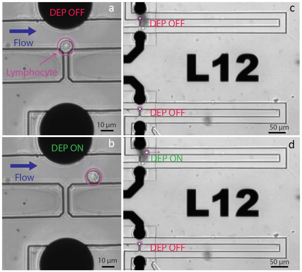
In another project, we enabled the capture of single cells using hydrodynamic traps. The device made it possible to release single cells selectively using dielectrophoresis, recover them and analyze their transcriptome by DropSeq [2]. We investigated the stress induced on cells by our platform through analysis of the molecular phenotype, revealing no impact of DEP application on the transcriptional signature of the cells. In general, this device could be used for controlled monitoring of single-cells that were subjected to different drugs or toxins.