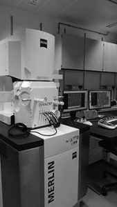
The microscope
The most recent addition to the facility is the Gatan 3view microscope. The microscope uses a combination of scanning electron micrscopy and an internal ultramicrotome to produce a series of images. With each section nm of resin are removed using the ultramicrotome and the new surface is then imaged. This allows the user to move through the sample and produce a detailed stack of information from which measurments can be taken and models can be made.
Last News:
A focal charge compensator was added to the microscope to delivers artefact-free images. When imaging resin embedded samples such as vascularised tissues, charging effect can appear.
How it works:
a tiny carbon needle is precisely located above the sample and rely Nitrogen gas directly onto the sample surface while the chamber maintains high vacuum. This allows the user to perform serial block face imaging with high acquisition rates and avoid imaging artefacts.