Cancer photodetection by fluorescence imaging is based on the administration of a tumor localizing fluorescing dye. Upon irradiation of the investigated area with excitation light, the dye will emit fluorescence light. In certain organs, the tissue native autofluorescence is bright and contrasted enough to allow the detection of superficial cancerous lesions. The two main clinical applications of this approach are in the tracheo-bronchial tree and the bladder.
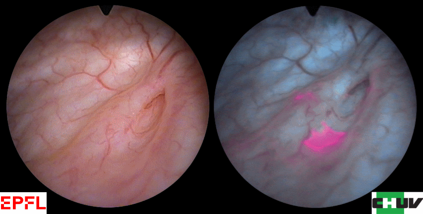
Photodetection of flat lesions (Carcinoma in-situ) in the bladder with white light (left) and ALA-Hexylester fluorescence cystoscopy (right).
The detection and quantitative removal of early bladder cancer by endoscopic fluorescence imaging represent a significant advance in the field of oncology. This procedure was partly developed by our group. It led to the products Hexvix® (Cysview® in the USA).
Although these detection approaches are quite sensitive, their specificity can be improved by optimizing the spectral design of the fluorescence imaging instruments or the diet of the patients: the fluorescence of the bladder washout fluid or the presence of hemoglobin in this fluid or inflammatory foci have a negative influence on the performance of this cancer detection procedure. Therefore, finding the optimal excitation and detection wavelengths for specific applications of cancer photodetection by fluorescence imaging as well as the identification of the optimal patient’s diet are not trivial and are some objectives of our group.
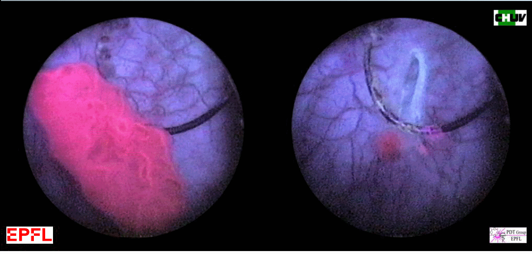
Fluorescence cystoscopy to monitor the resection of a bladder cancer
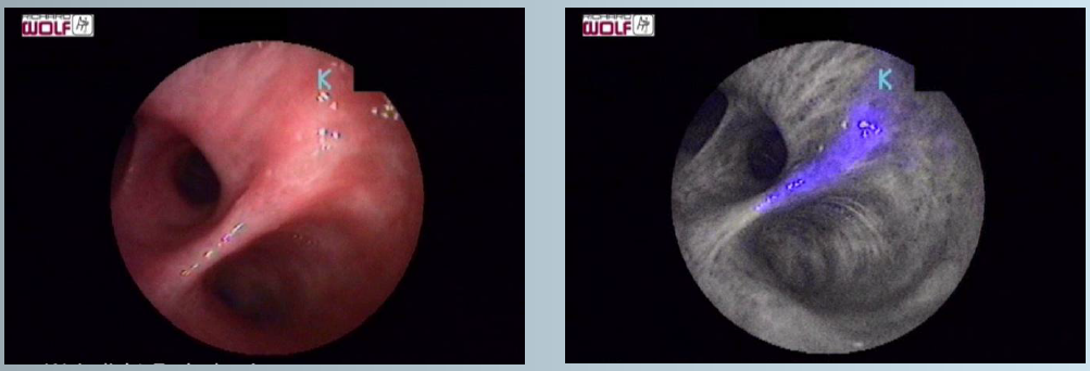
Detection of bronchial carcinoma in situ (CIS) by autofluorescence bronchoscopy
Below: Video examples of fluorescence cystoscopy for cancer detection/resection monitoring and autofluorescence bronchoscopy :