Understanding the functionality and layout of the human brain is a longstanding research goal in neuroscience. We develop methods to study the position and connection of neurons and other structures, for automatic segmentation and for tracking formations over extended periods of time.
Tracking Dividing Cells
Network Flow Integer Programming to Track Elliptical Cells in Time-Lapse SequencesWe propose a novel approach to automatically tracking elliptical cell populations in time-lapse image sequences. Given an initial segmentation, we account for partial occlusions and overlaps by generating an over-complete set of competing detection hypotheses. To this end, we fit ellipses to portions of the (…)
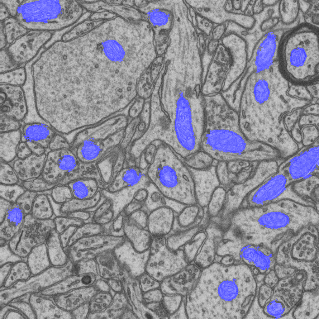
Mitochondria Detection in EM Stacks
click on the images to jump to some results.Electron microscopy (EM) is key to mapping the morphology of neural structures. Recent techniques, such as Focused Ion Beam Scanning Electron Microscopy (FIB-SEM), can now deliver image stacks at the nanometer resolution in all three dimensions. Such stacks show very fine structures that are critical to unlocking (…)
Synapse Detection in EM Stacks
Learning Context Cues for Synapse Segmentation in EM VolumesWe present a new approach for the automated segmentation of excitatory synapses in image stacks acquired by electron microscopy. We rely on a large set of image features specifically designed to take spatial context into account and train a classifier that can effectively utilize cues such as (…)
Automated Reconstruction of Complex Curvilinear Structures using Path Classifiers and Integer Programming
click on the images to jump to the corresponding results.Networks of curvilinear structures are pervasive both in natural and man-made systems. They appear at all possible scales, ranging from nanometers in Electron Microscopy images of neurons and meters in aerial images of roads to petameters in dark-matter arbors binding massive galaxy clusters.Current practical approaches for (…)
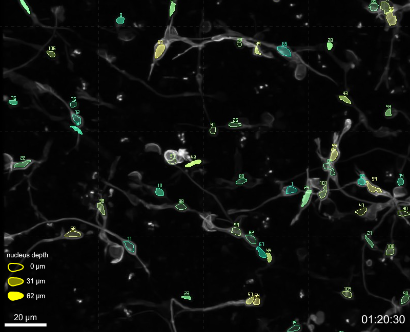
Detecting and Tracking Migrating Neurons
click on the images to jump to some resultsWhile most neurons are generated during the embryonic and postnatal periods, it is now well accepted that some regions of the brain keep producing new neurons throughout adulthood. Understanding the processes that govern the birth and development of neurons could lead to future treatments to a variety (…)
Non-Rigid Registration of Neuronal Stacks
click on the images to jump to some resultsElectron Microscopes can now provide the nanometer resolution that is needed to image synapses, and therefore connections, while Light Microscopes see at the micrometer resolution required to model the 3D structure of the dendritic network. Since both the arborescence and the connections are integral parts of the (…)