
Keywords: FD-FLIM, microscopy, modulation, scattering
Background
FLIMME is the first fluorescence lifetime endomicroscope combining wavefront shaping through a single multimode fiber (MMF) with frequency-domain fluorescence lifetime imaging (FD-FLIM) capable of imaging various samples with sub-micrometer spatial resolution and temporal precision of approximately 30 ps [1]. The system exploits the transmission matrix of the multimode fiber to generate diffraction-limited excitation at the distal tip while preserving the minimally invasive footprint of a needle-like fiber probe. Fluorescence lifetime provides endogenous biochemical and metabolic contrast, allowing discrimination of tissues without exogenous labels.
Several important challenges limit the imaging capabilities of FLIMME in dense, non-sparse fluorescent samples. In transmission-matrix wavefront shaping, the achievable enhancement and power ratio depend on the fraction of controlled optical modes, i.e. the available degrees of freedom (DOFs). The approximate number of guided modes in a step-index multimode fiber increases with increasing core diameters and NA. Since the wavelength and the number of DMD pixels are fixed by the system, increasing the number of supported modes reduces the fraction of controlled modes. Consequently, probe design requires balancing field of view (core diameter), spatial resolution (NA), and imaging contrast (enhancement and power ratio).
A second challenge concerns volumetric imaging. Two-dimensional reconstructions of a probing event require accurate compensation for motion artifacts originating both from sample deformation during insertion and from the mismatch between line-scanning speed and translation-stage motion. Finally, the point spread function varies across the scanning field in transmission-matrix imaging, requiring dedicated post-processing to obtain quantitative 3D reconstructions.
Finally, validation against histological ground truth presents practical challenges. Correlating fluorescence lifetime images with cryosectioned tissue requires sectioning along the probe insertion path after imaging. While this process is relatively straightforward for a single insertion, multiple probe insertions can cause tissue displacement during probe extraction, making it difficult to recover the exact imaging locations. Appropriate probing strategies and registration methods therefore need to be developed to ensure accurate comparison between endomicroscopy and histopathology.
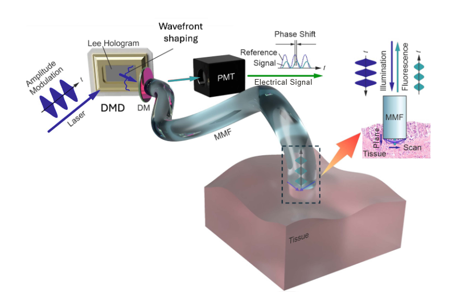
Figure 1 – Principle of FLIMME
Video description of the project’s context and description on the ISREC’s website
Project description
The objective of this project is to optimize the FLIMME platform for imaging a range of biological samples, including organoids, transwell cultures, and excised tissue specimens.
For each sample type, the student will determine the optimal imaging conditions by investigating different multimode fiber probe configurations and insertion parameters. This includes selecting appropriate probe geometries, insertion speeds, and acquisition settings according to the mechanical properties of the sample (e.g., viscosity, density, stiffness) and the biological feature size that must be resolved. Experimental data will be acquired and quantitatively analyzed to establish standardized acquisition protocols for each sample type.
The student will investigate image enhancement strategies through both hardware-aware preprocessing and computational post-processing. This includes developing methods to compensate for motion artifacts, correcting position estimation during volumetric acquisitions, accounting for the position-dependent point spread function, and exploring reconstruction algorithms that improve image quality and quantitative fluorescence lifetime measurements.
Finally, the student will design and fabricate sample holders adapted to the different biological models and develop an automated insertion and imaging strategy using the motorized translation stage. The resulting workflow should enable reproducible volumetric acquisitions while minimizing tissue deformation and improving correlation with subsequent histological analysis.
Throughout the project, the student will gain experience in advanced optical microscopy, fluorescence lifetime imaging, wavefront shaping through multimode fibers, automated instrumentation, image processing, and quantitative analysis of biological imaging data.
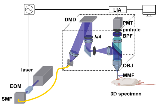
Figure 2 – Schematic of the current FLIMME optical setup
Student profile
The student should be interested in imaging techniques and biology. The student should be motivated and organized. They should have good communications skills in English as the report shall be written in that language. The student should have some experience with Matlab/Python. Previous experience with fluorescence microscopy and optics is appreciated.
Contact
If you are interested in this project, please contact me by email at: [email protected]
References
[1] Fay et al. arXiv:2510.00267