Imaging with multimode fibers

When images pass through optical fibers they become scrambled due to the different propagation velocities of their spatial modes. As a result of this, the different modes interfere on exiting from the fiber to produce a specular pattern. Research in the optics laboratory has focused on recovering the input images from their corresponding speckle patterns using deep neural networks. In particular, we have studied the effects of wavelength drift, and the difference between intensity only and holographic recording of the speckles on the reconstruction results. Work has also being carried out on reconstructing images obtained through multicore optical fibers using deep-learning techniques for the development of novel endoscopic microscopes.
Publications :
- Learning to see through multimode fibers
N Borhani, E Kakkava, C Moser, D Psaltis
Optica, 5(8):960-966 (2018) - Imaging through multimode fibers using deep learning: The effects of intensity versus holographic recording of the speckle pattern
E Kakkava, B Rahmani, N Borhani, U Teğin, D Loterie, G Konstantinou, C Moser, D Psaltis
Optical Fiber Technology 52, 101985 (2019) - Deep learning-based image classification through a multimode fiber in the presence of wavelength drift
E Kakkava, N Borhani, B Rahmani, U Teğin, C Moser, D Psaltis
Applied Sciences 10 (11), 3816 (2020)
Optical diffraction tomography
Solving the missing cone problem in optical diffraction tomography (ODT)
Optical diffraction tomography (ODT) provides us with the 3D refractive index (RI) distributions of transparent samples by acquiring 2D holograms from multiple angles. Inverse models are able to retrieve the 3D distributions from those 2D projections. However, the final reconstruction suffer from what is called “missing cone problem” due to the limited numerical aperture (NA) of the imaging system. In our group, we aim to solve this problem by 2 approaches. The first is a model based iterative reconstruction scheme. The second approach is based on neural networks.
Intensity-only optical diffraction tomography (ODT)
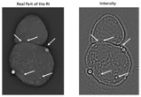
Instead of using holographic data to reconstruct the 3D RI distribution of the sample using inverse models, inverse models were directly applied on 2D intensity projections. Although the final reconstruction does not explicitly represent the 3D RI distribution of the samples, the high frequency features of the sample were more pronounced as compared to 3D reconstructions using 2D holographic data acquired from different projections. This property can be used to localize nanoparticles such as lipids inside a biological sample. In one of our collaborations we used these reconstructions to investigate the presence of nanoparticles on the membrane of immune T-cells.
Polarization sensitive optical diffraction tomography (ODT)
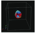
Polarization of light has been widely used as a contrast mechanism in many imaging modalities. In our group we reformulated inverse models to reconstruct the RI tensor of the samples which is directly linked to the birefringence of the sample. The off-diagonal components of the RI tensor reconstruction convey additional information that is not available in either conventional scalar ODT or 2D polarization microscopy. We used those vector-based models to estimate the birefringence of biological samples including human muscle tissues.
Publications :
- Polarization-sensitive optical diffraction tomography
A. Saba, J. Lim, A. B. Ayoub, E. E. Antoine, D. Psaltis
Optica 8, 402-408 (2021) - 3D reconstruction of weakly scattering objects from 2D intensity-only measurements using the Wolf transform
A. B. Ayoub, J. Lim, E. E. Antoine, D. Psaltis
Opt. Express 29, 3976-3984 (2021) - Fluorescence-Based and Fluorescent Label-Free Characterization of Polymer Nanoparticle Decorated T Cells
T. Thomsen, A. B. Ayoub, D. Psaltis, HA Klok
Biomacromolecules2021 22 (1), 190-200, DOI:10.1021/acs.biomac.0c00969 - Deep learning approach for solving the missing cone problem in optical diffraction tomography
J. Lim, A. B. Ayoub, and D. Psaltis
Imaging and Applied Optics Congress, OSA Technical Digest (Optical Society of America, 2020), paper CF4C.5. - Optical Diffraction Tomography (ODT) for Label-Free Imaging of Large 3D Biological Samples
E. E. Antoine, J. Lim, A. B. Ayoub, N. Brandenberg, and D. Psaltis
Imaging and Applied Optics Congress, The Optical Society (Optical Society of America, 2020), paper HF1G.2. - Three-Dimensional Optical Diffraction Tomography With Lippmann-Schwinger Model
T. -a. Pham, E. Soubies, A. Ayoub, J. Lim, D. Psaltis and M. Unser
IEEE Transactions on Computational Imaging, vol. 6, pp. 727-738, 2020, doi: 10.1109/TCI.2020.2969070. - A method for assessing the fidelity of optical diffraction tomography reconstruction methods using structured illumination
A. B. Ayoub, T. Pham, J. Lim, M. Unser, D.Psaltis
Optics Communications, Volume 454, 2020, 124486, ISSN 0030-4018. - High-fidelity optical diffraction tomography of multiple scattering samples
J. Lim, A.B. Ayoub, E.E. Antoine, D.Psaltis
Light Sci Appl 8, 82 (2019). - Predicting optical transmission through complex scattering media from reflection patterns with deep neural networks
K Skarsoulis, E Kakkava, D Psaltis
Optics Communications, Volume 492, 2021, 126968, ISSN 0030-4018