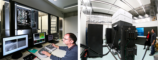
CIME Day 2020 6 March, Rolex Forum
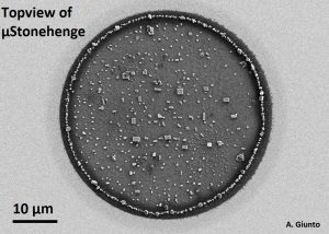
best picture: Andrea Giunto
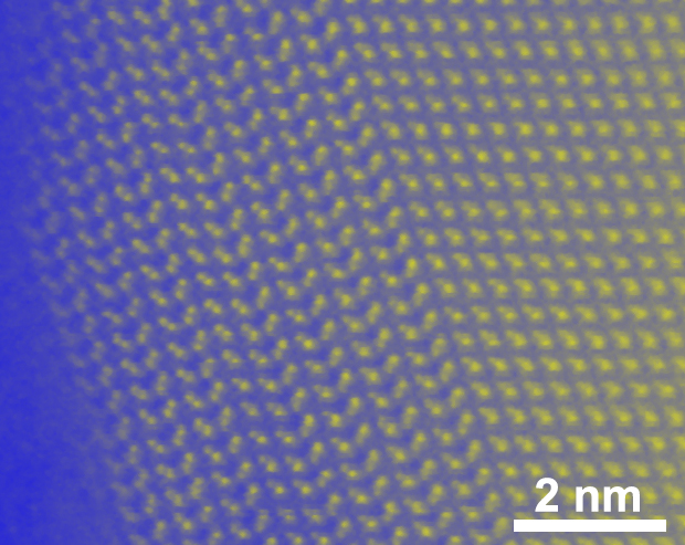
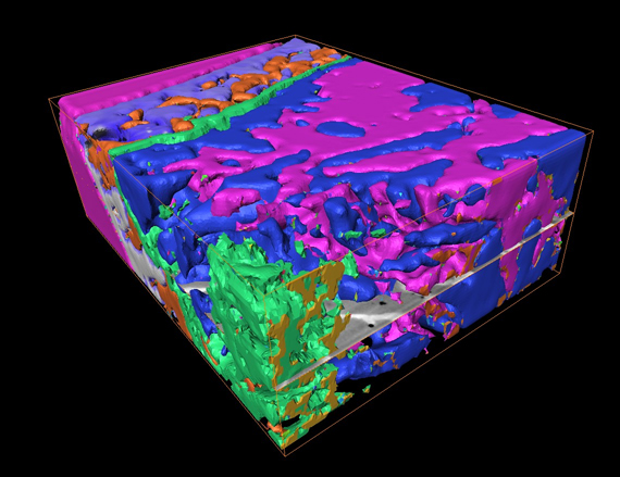
best Life-Science: Nathan RIGUET, SV-LMNN
Nathan RIGUET Poster CIME2020 Final
best poster Materials Science: Fabien GEORGET, STI-IMX-LMC
Seminar announcement:
Seminar announcement: Friday 29th of November, MXC-315
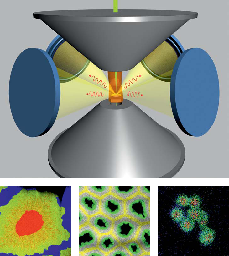
eCHORD a method of mapping orientations with high resolution in SEM
Dr. Cyril Langlois, Laboratoire MATEIS INSA Lyon
The “Microscopies” team of the MATEIS laboratory (INSA Lyon) has developed a new method of mapping orientations and phases based on an electron-matter interaction that is starting to be exploited thanks to the improvement of the detectors and processing computers in the last decades. This is the electron channeling by the crystalline planes, a phenomenon that explains the contrast between the grains on an electron microscopy image. In this talk, the principle of using the channeling phenomenon to go back to the crystalline orientation (eCHORD approach) will be explained. The main advantages of the eCHORD technique are a better spatial resolution thanks to the low voltage (possible mappings down to 1 kilovolt instead of the usual 10kV in EBSD), and simplicity of use (simplified acquisition geometry, no additional detector in the microscope). The performances of the technique will be put forward, especially in comparison with the EBSD.
New online cours on transmission electron microscopy
Transmission Electron Microscopy for Materials Science new on Coursera: https://www.coursera.org/learn/microscopy
Built from the ground up, the course is intended to be a starting point for understanding and working with TEM; to be used either as a stand alone resource, or in complement to books and in person teaching or coaching. We hope that it proves useful to our electron microscopy community, and look forward to receiving any feedback.
With regards
Cécile Hébert and Duncan Alexander
EPFL-SB-IPHYS-LSME
Congratulations !
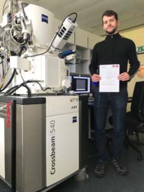
Emad Oveisi wins Microscopy Today Innovation Award
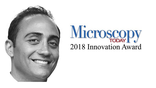
see EPFL Memento
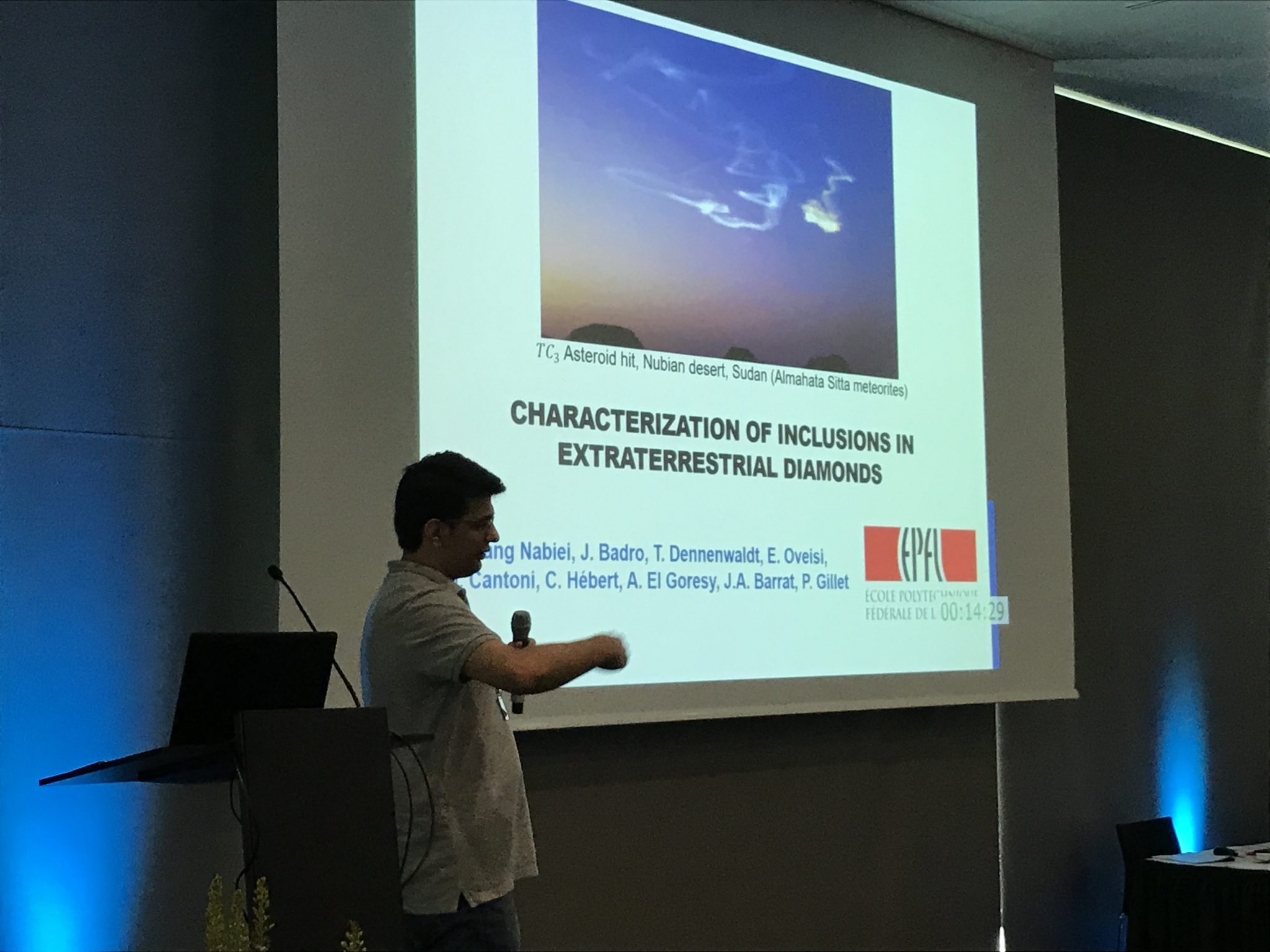
“Diamonds from a lost planet”
Congratulations Farhang Nabiei for your Nature Communications paper (link here ) that produced such a refreshing excitement in the news (see rdp.epfl.ch of april 18th for example).
Here a souvenir picture of your thesis defense presenting a “fancy” story…
CIME Day 2018
CIME-Day 2018, general assembly and user’s day
…. and the poster prices go to
Urszula Cendroswska STI-IMX-SUNMIL for the poster:
Gold nanoparticles as a novel tool in amyloid studies
and
Stéphane Poitel SB-IPHYS-LSME for the poster
Electron Microscopy : From In-Situ Observations to Post-Test Microstructure and chemical analyses
Congratulations and Thank you !!
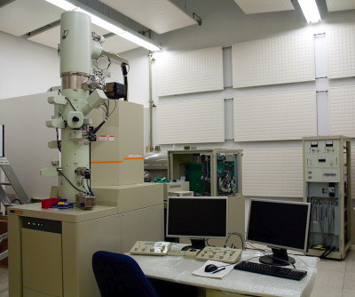
new Gemini-SEM 300 operational !
Dear XLF-30 users,
The new CIME SEM (Zeiss GEMINI 300) is installed, and we are planning the training/transition of the XLF-30 users on the new instrument.
We offer all trained XLF-30 users, a ½ day basic training/transition on the new Gemini-SEM (free of charge). The EDX-training (new EDX System and interface) will not be covered in this transition and requires a separate training (free of charge for users already trained for EDX).
The advanced and new options available on this instrument also require a separate training request and will be charged according to the new rules, i.e., users will be billed for the microscope hours and operator-assisted service.
To schedule your basic and advanced training sessions, we will ask that you fill the form below and send it to [email protected] as soon as possible.
MC2017 news
Congratulations to conference chairs Marco Cantoni, Rolf Erni and Markus Dürrenberger for a successful Microscopy Conference 2017 held in the SwissTech Convention Center from 21–25 August!
In the MC2017 awards, congratulations to:
- Pelin Güven of SUNMIL for winning a student poster prize in the session MS 5 on Carbon-based materials, soft matter and polymers for her poster entitled: Nanoparticles for modification of vesicles.
- Dr Emad Oveisi of CIME for taking 3rd place in the Best Image Contest “Art in Science 2017” for the image entitled: Rice Terraces – Faceted grain growth in hexagonal barium titanate ceramics:
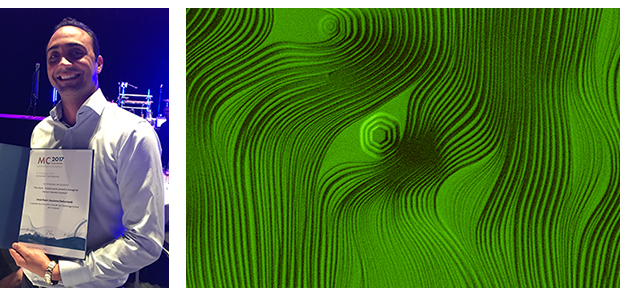 Width of barium titanate image: 24 μm.
Specimen courtesy of Sina Hashemizadeh of Ceramic Laboratory – EPFL.
Decommissioning of XLF-30 SEM
The days of the faithful XLF-30 are counted !
It will be replaced with a ZEISS Gemini-SEM 300 in August.
In order to ease the transition we have found an agreement with Prof. G. Fantner’s lab where the equipment will serve as an experimental platform in the future:
On July 11/12 the XFL-30 will be transferred to the BM building. Prof Fantner has agreed to make the instrument available to the CIME users until they have been trained on the new SEM.
You will be able to continue to reserve and use the XLF-30 in its new place as soon as it has been moved and tested. As soon as the new microscope will be fully operational we will start to train XLF-30 users on the new platform (free of charge).
MC2017
Atomic column imaging
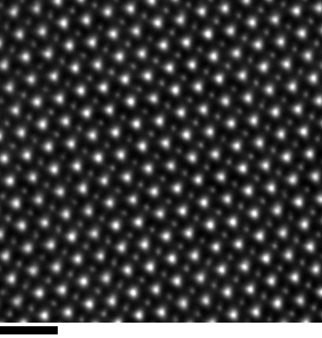
DyScO3 imaged by high angle annular dark field scanning TEM on the Titan Themis 60-300. The heavier Dy cation columns, showing as brighter spots, displace in alternate directions relative to the the lighter Sc cations. Scale bar: 1 nm
Image: Duncan Alexander
Specimen preparation: Danièle Laub and Colette Valloton
Sample material courtesy of the Triscone Group, UNIGE
CIME Day 2016
Dear colleagues,
On Monday October 17 we will hold CIME Day: the first in a series of annual meetings of the Interdisciplinary Center for Electron Microscopy (CIME), EPFL. In this meeting we are excited to host speakers from EPFL and beyond, who will present on many different areas of electron microscopy (see schedule below). It will take place at the Rolex Learning Center from 9am to 3pm, with lunch and coffee breaks included, and will provide a forum for networking and discussion to all users.
All CIME users are invited to register for this event. We encourage each attendee (PhD students, postdocs and collaborators) to present a poster summarizing the use of electron microscopy at CIME in their research. The presenter of the best poster will be awarded a prize of 100 CHF and his/her lab will be given one free user training at CIME.
Registration is required by sending an email to: [email protected]
The deadline for registration is September 26 at 5pm.
Cordially
Cécile Hebert, Marco Cantoni
EPFL SB-CIME, Batiment MXC 132
Station 12, 1015 LAUSANNE, Switzerland
For the CIME’s Executive Committee
Anna Fontcuberta i Morral
Program:
9:00 Welcome
9:10 Orientation imaging microscopy: quantitatively tracking the crystal
grain structure down to the nanometer scale, Brian Aebersold
9:30 Cryo-electron microscopy and tomography, Paul Guichard
9:50 Three-dimensional focused ion beam for life and materials sciences Marco Cantoni
10:10 Block face imaging in life sciences, as a complementary tool to 3D-FIB, Catherine Maclachlan
10:30 coffee break and poster session
11:00 Cathodoluminescence spectroscopy, David Gachet
11:20 Challenges and perspectives in aberration corrected (S)TEM, Sorin Lazar
11:50 high throughput STEM and STEM-EDX, the point of view of a user, Florent Héroguel
12:05 lunch break and poster session
13:15 Electron spectroscopies in the TEM for understanding nano structures and vice-versa, Odile Stéphan
14:00 coffee break and poster session
15:00 poster prize and closing remarks
Titan Themis Inauguration
CIME is pleased to announce the inauguration ceremony of the FEI Titan Themis on Thursday 1st and Friday 2nd October, 2015, at EPFL. The two-day ceremony will feature a series of scientifc seminars by world leading microscopy experts.
For full details of the seminar programme and other inauguration ceremony details please click here.
The sub-Å era arrives at CIME EPFL
CIME is pleased to announce that the installation of EPFL’s double Cs-corrected, monochromated FEI Titan Themis is completed, with the instrument performing well within specification. CIME staff are now working to take first application data on a variety of samples. To help illustrate the capabilities of the microscope the image shows a hexagonal phase wurtzite inclusion at the edge of a cubic phase zinc blende (Al,Ga)As shell on a GaAs nanowire core. Taken in STEM mode using the “Z-contrast” HAADF detector at 200 keV beam energy, a probe of <0.8 Å resolution allows the individual atomic columns in the (Al,Ga)As “dumbells” – separated by just 1.4 Å – to be clearly resolved across the defect interface.
Image by Duncan Alexander, CIME. Sample kindly provided by Yannick Fontana and Anna Fontcuberta i Morral, LMSC. False colour added for clarity.
Zeiss Merlin Calibration Problem 10.10.2014-16.1.2015
After the installation of a SmartSem software patch the output device and image calibration was corrupt.
The following description explains how to recover the right image calibration.
The tools mentioned in the description can be found below.
If you encounter problems, don’t hesitate to come to our office hours.
Marco Cantoni
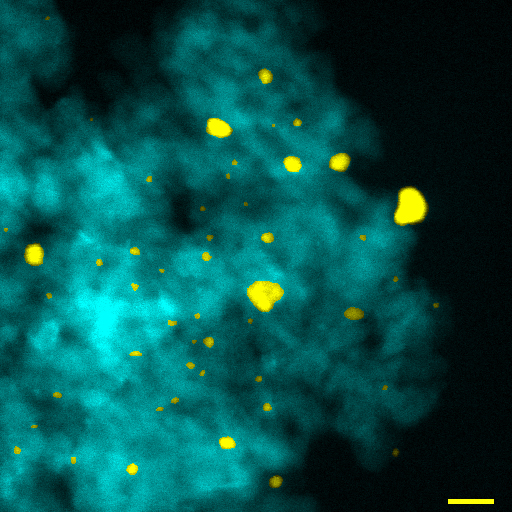
CIME BioEM – Nature Neuroscience cover
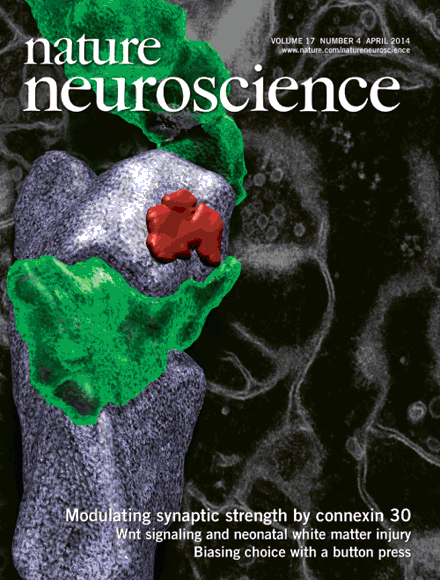
Cover image article in Nature Neuroscience. This month’s cover of Nature Neuroscience shows a 3D rendering of a dendritic spine reconstructed from serial electron micrographs from the hippocampus of an adult mouse. The image relates to collaborative research between the group of Natalie Rouach in Paris and Graham Knott in the bioEM Facility. This study shows how the protein CX30 (connexin 30) regulates synaptic transmission by controlling the extension of astrocytic processes into the synaptic cleft.
Click here to go to the journal article.
Nanoscale grain mapping
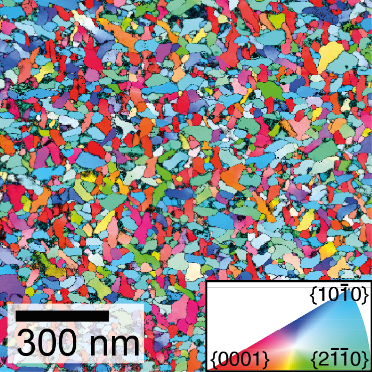
Using the NanoMEGAS ASTAR system installed on the JEOL 2200FS TEM instrument, the size and orientation of grains in a polycrystalline ZnO thin film are mapped with a 2 nm step size. In this way grains with diameters of <20 nm are identified, far below the limit offered by conventional SEM-based EBSD. The map data are presented as an inverse pole figure, with a colour coding made with respect to the East direction, and with more reliable pixels illustrated as brighter colours. The ZnO film was supplied by Lorenzo Fanni, Aïcha Hessler-Wyser and Christophe Ballif of the PV-lab and Sylvain Nicolay of the CSEM’s PV-center, within the framework of the ZONEM project.
A. Brian Aebersold, Duncan Alexander and Cécile Hébert, CIME, EPFL.
New Doctoral School format
CIME is pleased to announce the introduction of a new modular doctoral school format for 2014, which will allow PhD students and researchers to select the components most relevant to their needs. Starting in the spring 2014 semester we offer the following doctoral schools:
Scanning Electron Microscopy Techniques
Transmission Electron Microscopy and Diffraction
Scanning and Analytical Transmission Electron Microscopy
3D Electron Microscopy and FIB-Nanotomography
Electron Microscopy for Life Science
If you have any questions about the courses please consult their webpages or contact the CIME team by emailing [email protected] or visiting us during CIME office hours (Tuesdays and Fridays 08h30-10h00 MXC 133).
CIME Office hours
To aid communication with EPFL researchers CIME is introducing a system of “office hours” every Tuesday and Friday morning from 08h30 to 10h00, MXC 133 where you can meet with one of CIME’s scientific staff. During these periods researchers working on physical/materials systems are encouraged to come and discuss questions related to use of electron microscopy for their projects, for instance:
- training and service requests
- selection of electron microscocopy techniques
- TEM and SEM results
- project feasibility
Super-X User Meeting 2014
Together with the support of FEI, CIME is pleased to announce the Super-X User meeting taking place at EPFL, Lausanne on the 13-14 Febuary 2014. The meeting will focus on the Super-X EDX system of the FEI Tecnai Osiris, Thalos and Titan ChemiSTEM instruments and its specificities. It will give Super-X users the opportunity to present and discuss their scientific results and technical challenges and solutions. The meeting webpage and program can be found here.
CIME researchers rewarded by EPFL
Marco Cantoni has been awarded the prize of the Fondation Rodolphe et Renée Haenny, recognizing his “strong commitment in innovating and promoting electron microscopy tools in the field of materials science, but also in other disciplines such as basic and life sciences. His involvement in the development of sophisticated 3-dimensional analysis techniques, in particular based on focused-ion beam, has placed the Interdisciplinary Centre of Electron Microscopy of EPFL as one of the world-leading groups in this area”.
J. Andreas Schuler‘s PhD thesis “Chromium Poisoning: The Needle in the SOFC Stack” has been awarded the Professor René Wasserman Award 2013. The prize, for contributions of high value to the theory and application of materials science, recognized the thesis for its “comprehensive insight into slow incipient deactivation of catalysts by volatile traces, their quantification, localisation and avoidance”. Dr Schuler, who made his PhD at CIME, is now a project manager for fuel cell specialists Hexis AG.
Brain axons and dendrites in 3D
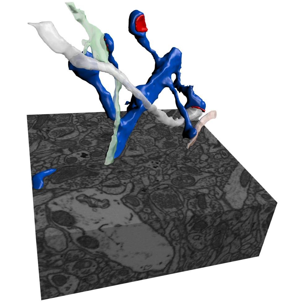
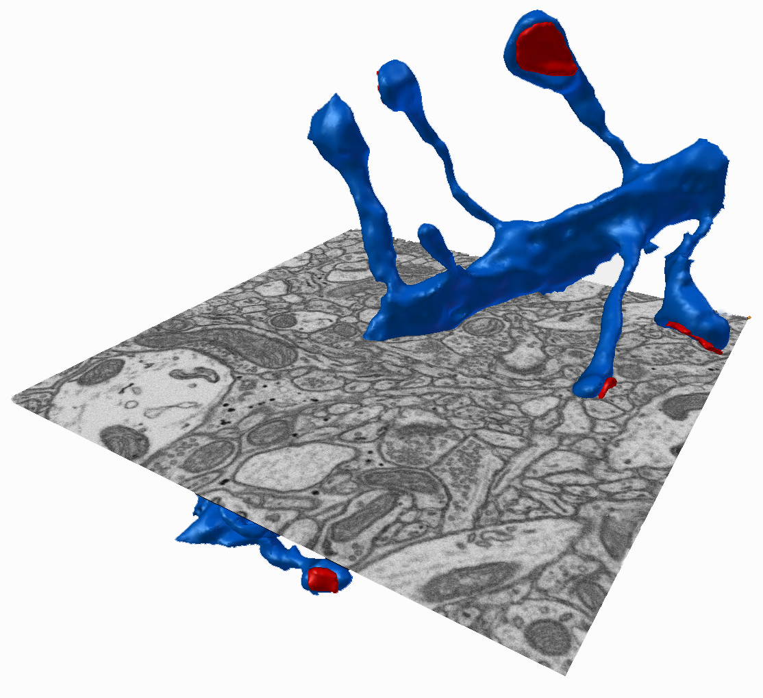
The images show axons (white) and dendrites (blue) within a volume of adult mouse brain tissue imaged with FIBSEM. They were reconstructed automatically with the Ilastik software, and analysed and rendered within the Blender software. Also shown are the synaptic connections, in red, situated on the end of the dendritic spines.
Courtesy of Corrado Cali, FIBSEM by Marco Cantoni, CIME.
Mycobacterium tuberculosis
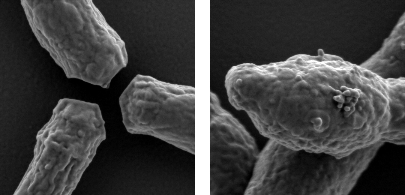
Images of Mycobacterium tuberculosis taken with the new Zeiss Merlin SEM. On the left is the H37Rv wild type displaying a normal bacillus-like shape. In contrast, on the right are H37Rv mutants lacking an enzyme involved in the cell wall biosynthesis, which leads to the cell swelling observed.
Image conditions: 1 kV beam energy; 2 mm working distance; in-lens SE detector. Each image shows a 1.2 x 1.2 μm region.
Images kindly provided by Gaëlle Kolly of the Cole Lab and Fabienne Bobard of CIME.
Happy new year 2013
CIME thanks all users for collaborating in 2012, and wishes them a happy and productive year in 2013.
GaAs nanowire with Ga droplet. EDX: Tecnai OSIRIS chemistem. Analysis by Sonia Conesa Boj. Image compressed in x.
Sample: A. Fontcuberta i Morral, LMSC
Red: Si; blue: As; green: Ga
Flux vortices imaged in MgB2 superconductor
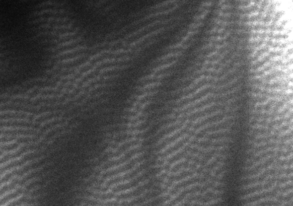
A lattice of flux vortices in MgB2 is successfully induced and imaged using Lorentz mode microscopy in the JEOL 2200FS TEM, a liquid helium stage cooling the sample to 5 K, and a specimen prepared using the Zeiss NVision 40 FIB. In this image the applied magnetic field was tuned to give a vortex separation of about 200 nm.
Image and experiments by Mathieu Cottet and Barbora Mansart of Fabrizio Carbone’s LUMES laboratory, and Marco Cantoni and Duncan Alexander of CIME.
“Super-X” EDX mapping on Osiris – Fe/Al friction weld
512 x 512 px elemental map of a friction weld between aluminium alloy and carbon steel, taken in only ~10 minutes using the FEI Tecnai Osiris TEM with Super-X EDX acquisition system.
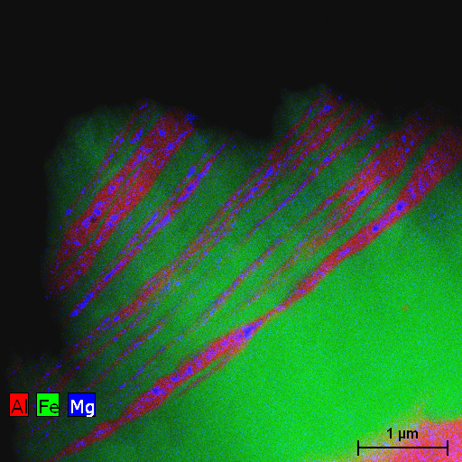
Sample: Friction stir lap welded Al alloy and carbon steel. During welding, layered structures form near the joint interface in different micro- and nano-scales because of stirring and mixing effects, with aluminum layers entering into the steel transforming to an Al/Fe intermetallic compound under high temperature. Mg in the Al alloy is rejected by the intermetallic, segregating to form inclusions along the centerlines of the layers.
Sample and description provided by H. Najafi (ICMP, EPFL) in collaboration with M. Movahedi, A.H. Kokabi and S.M. Seyed Reihani of the Department of Materials Science and Engineering, Sharif University of Technology.
Image by the CIME team.
FEI Tecnai Osiris installation near complete!
The CIME team is pleased to announce that the FEI Tecnai Osiris installation is near finished, and it should soon be entering regular service. Early results demonstrating the outstanding EDX analytical and mapping capability to be shown here shortly…
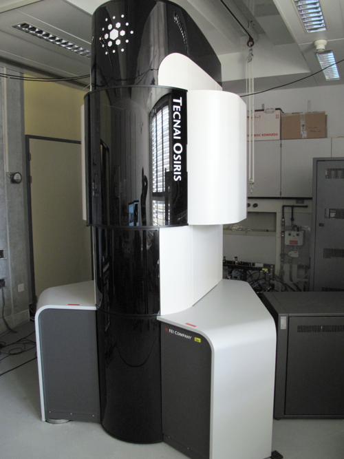
Autumn 2011 – New Tecnai Osiris now under installation
Embed of video is only possible from Mediaspace, Vimeo or Youtube
Image of the moment September 2011 – new PV technology
This month in Nature Photonics, Battaglia et al. use the stunning cross-section imaging ability of the Zeiss NVision FIB/SEM to demonstrate their nanomoulding technology in thin-film silicon photovoltaic cells.

Samples: Solar cells on master ZnO (left), nanomoulded ZnO replica (middle), and the nanomoulded ATA replica (right), Corsin Battaglia, IMT PV-lab
Images: Duncan Alexander, CIME
Coming soon – new microscopes at CIME!
Dear CIME users,
Three new microscopes will be installed in a short time frame starting from the end of August.
There will be some impact on the availability of machines and techniques during the microscopes’ installation and testing and until the users have been trained on the new machines.
The following microscopes will be stopped:
- CM20: third week of August
- CM10: end of September
Installation of new microscopes:
- First week of september: Tecnai Spirit Bio Twin: basic Bio-EM
- End of September: Tecnai G2 F20 “Cryo-Bio-Tomo” (will free capacity on the cm12 and JEOL)
- Beginning of October: Tecnai OSIRIS (high throughput analytical TEM for Materials Science)
Impact on training and availability of machines:
- Training and use of basic Bio-EM (until now CM10) might be interrupted for 1-2 weeks.
- Basic TEM training for Materials science (CM20 and CM12 until now):
o New users for basic TEM will be trained on the CM12 until end of the year.
o Regular CM20 users continue basic TEM on the CM12. Transition (if not yet done) takes less than 30min “training” (free of cost). Contact Aïcha Hessler-Wyser ([email protected]) for access rights and short training (if not yet done).
o Training on the OSIRIS: user training (experienced users and new users) will start after the acceptance tests and CIME staff training: Expected ~December/January
Details to be announced.
o CM10 users will get an introduction on the Tecnai Spirit in September. Details will be announced as soon as the microscope has been installed and accepted.
Thank you for your support
Marco Cantoni
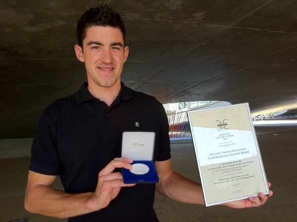
Bravo to Andreas Schuler!
Congratulations to J. Andreas Schuler et al., awarded the “Christian Friedrich Schönbein Contribution to Science Medal” by the committee of the European Fuel Cell Forum 2011 for their oral/proceeding contribution entitled:
Nd-nickelate SOFC Cathode Sensitivity to Cr and Si Contamination
by J. Andreas Schuler, Henning Lübbe, Aïcha Hessler-Wyser and Jan Van herle.
Their award-winning research is illustrated in the following figure:
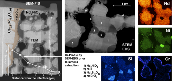
Left: SEM imaging of a TEM-lamella, extracted by FIB from the cell described below, showing an Nd2NiO4 (NNO) cathode region on a (Ce,Gd)O2 electrolyte support with an intermediate NdCeO3,5 reaction layer.
Right: annular dark-field STEM image and corresponding EDS elemental maps revealing Cr deposits on active NNO grain surfaces as well as secondary Ni- and Si-rich phases.
Details: Nd-nickelate cathode polarized under 0.5 A/cm2 current density during 1000 hours at 700°C in air with deliberate Cr contamination (J.A. Schuler and H. Lübbe). TEM-lamella preparation by FIB Zeiss Nvision40 (F. Bobard). EDS observations with SEM XLF-30 (J.A. Schuler). STEM-EDS investigation on TEM CM300 (A. Hessler-Wyser).
Bravo!
Congratulations to Pierre Burdet et al., awarded the 1st Prize by the committee of the European Microbeam Analysis Society 2011 Workshop for their Young Scientist’s contribution entitled:
3D EDS microanalysis by FIB-SEM: enhancement of elemental quantification
by P. Burdet, C. Hébert and M. Cantoni.
Picture of the moment – March 2011: 3D EDX
3D EDX microanalysis by FIB/SEM: Molten region of NiTi and stainless steel welding by laser. The stack of EDX maps is used to identify the different intermetallic phases and to reconstruct their microstructure in 3D.
Details: Acquisition using a FIB/SEM dual-beam Zeiss Nvision40. 44 EDX maps recorded with voxel size of 100x100x100 nm (126×92 pixels per map). In parallel, 352 in-Lens SE images recorded with voxel size of 12.5×12.5×12.5 nm. Sample of J. Vannod, LSMX.
These results have been presented during the 5th EDMX day. The presentation won the best presentation award.
P. Burdet, CIME
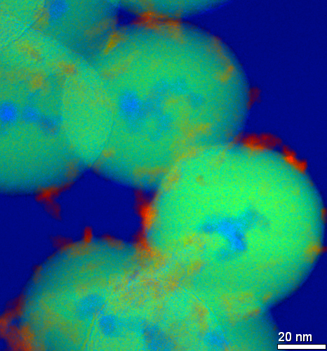
Picture of the moment – February 2011
The atomic number contrast of high angle annular dark-field scanning TEM imaging allows discrepancy of catalytic gold particles, down to < 1 nm in size, on an alumina support. Scale bar: 10 nm. False colour applied to highlight gold nanoparticles.
Details: The sample presented is a 1% wt. Au supported on alumina prepared by deposition-precipitation. Catalyst prepared by F. Cardenas-Lizana, I. Yuranov and L. Kiwi-Minsker, SB-ISIC-GGRC. Project title: “Rational Design of a Highly Active/Selective Structured Catalyst for Three-Phase Hydrogenations in a Compact Reactor”. Financed by Swiss National Science Foundation (200021-118067 1)
D.T.L. Alexander, CIME.
Picture of the moment – October 2010
Energy-filtered TEM RGB image of coated nanoparticles; titania: red; silica: green; iron oxide particle cores and vacuum: blue. D.T.L. Alexander, CIME.
Sample produced by U. Sakulkhu and H. Hofmann, LTP.
Project title: Preparation of coated nanoparticles and investigation of their behavior in biological fluids.
Financed by Swiss National Science Foundation (FN205321-120161).
Mai 2009: Cathodoluminescens installed and working
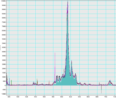 The mono-CL detection system has been succesfuly installed on the Jeol. Beautiful CL-spectra of YGdO nanoparticles shows the nice resolution of the system.
January 2009 : New Jeol arriving
 The new TEM Jeol 2200 FS is currently under installation. The FEG has just be installed
December 2008: SEM image
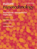 SEM image taken at CIME on the cover of Nature Nanotechnology, from the article “Borosilicate nanoparticles prepared by exothermic phase separation” by V.K. Parashar et al.