Partner: FORTH
Operator: Georgios Theodoropoulos LAPI/EPFL, CSTACC/FORTH, ([email protected])
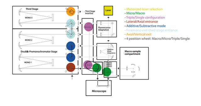
For the chemical and structural characterization of the samples, Raman spectroscopy was carried out using a NIR–Vis micro/macro-Raman system with the HORIBA Jobin Yvon T-64000 spectrometer. In the microscope (micro-Raman) configuration, a halogen lamp illuminated the sample surface, allowing precise selection of the measurement spot and visual inspection both before and after laser exposure.
To excite the Raman signal, three laser lines were used according to experimental requirements:
- 785 nm (CrystaLaser diode laser), maximum power ~7 mW
- 632.8 nm (He–Ne laser, Optronics Technologies S.A., model HLA-20P), maximum power ~3.5 mW
- 514.5 nm (Ar⁺ laser, Spectra Physics, water-cooled), maximum power ~10 mW
The choice of excitation wavelength was carefully optimized to minimize sample fluorescence and prevent laser-induced damage. Prior testing and adjustment ensured high-quality Raman spectra.
Before reaching the sample, each laser beam passed through a narrowband interference filter, which removes secondary emission lines from the laser. In the microscope setup, the beam was directed onto the sample, and the scattered light was collected through a beam splitter and the objective lens. Notch or edge filters blocked Rayleigh elastic scattering before the signal entered the spectrometer, allowing only Raman photons to be analyzed.
The scattered light was then directed into the monochromator, where a diffraction grating (either 1800 grooves/mm or 600 grooves/mm) dispersed the light according to wavelength. Detection was performed with a Symphony II 2D CCD detector (1024 × 256 pixels), cooled with liquid nitrogen to −133 °C to minimize thermal noise. Measurements were carried out using one of three objective lenses—10×, 50×, or 100×—depending on the desired spatial resolution.