
Our lab develops advanced acquisition and reconstruction methods for body MRI across mid- to high-field systems (0.55–3T), spanning pulse sequence design, reconstruction methods and parameter mapping. Student projects offer hands-on work at the interface of MRI physics, signal processing, and computational methods spanning model-based reconstruction and machine learning.
Master Thesis projects
Relaxometry on the transverse plane (T2) and in the rotating frame (T1ρ) play an important role in the study of brain pathologies as well as cartilage osteoarthritis. However, conventional spin-echo-based sequences for T2/ T1ρ mapping are limited to 2D or 3D-slab imaging with anisotropic resolutions due to high energy deposition and long acquisition time. In this project, we investigate magnetization preparation approaches (T2 or T1ρ) combined with fast gradient echo sequences that allow for volumetric and isotropic high-resolution mapping of the entire organ of interest.
Our current technique employs incoherent undersampled data acquisition and compressed sensing (CS) image reconstruction. To further reduce acquisition times and/or improve robustness and signal-to-noise, we have recently implemented a deep learning (DL) framework for reconstruction. The network has been trained on different gradient echo images of the brain and finetuned at different field strengths. This already showed improved SNR of knee T1rho maps at 3 T and promises to reduce acquisition times by few minutes, making the technique more suitable for clinical workflow.
Goals:
- To characterize the performance of the current DL framework in comparison to CS in a standardized phantom (ISMRM-NIST) and in healthy subjects (e.g. linear regression and Bland-Altman analysis of T2/T1rho estimates).
- To finetune the current DL model with QuantoRAGE images of knee and hip cartilage.
- To characterize the finetuned DL model in comparison to current DL model and CS in phantom and healthy subjects.
- (Optional) To investigate other accelerated acquisition strategies (GRAPPA/CAIPIRINHA) in combination with DL reconstruction.
The student will have the opportunity to:
- Learn about CS and DL reconstructions
- Learn about acceleration strategies for acquisition
- Have access to source DL recon for finetuning
- Acquire MR data in phantoms and in humans
- Perform comparison analysis of different methods (statistics)
Requirements:
- Knowledge of signal and image processing.
- Basic knowledge of MR physics.
- Basic knowledge of Machine Learning.
- MATLAB, Python programming. C/C++ is an advantage.
- Independent worker / Problem solving attitude
- Good command of English.
Duration: 6 months
Supervisors:
Day-to-day supervision and location: Gabriele Bonanno, PhD , Translational Imaging Center of sitem-insel, Bern
Academic supervisors: Jonathan Stelter, Prof. Dimitrios Karampinos
The project will be carried out in close collaboration with the Acquisition and Reconstruction groups of the Swiss Innovation Hub, Siemens Healthineers, Lausanne.
How to apply: please send your CV and cover letter to [email protected]

References:
- Bonanno G, et al. Program number 1475. Intl. Soc. Mag. Reson. Med. 29 (2021)
https://cds.ismrm.org/protected/21MPresentations/abstracts/1475.html - Forman C, et al. High-resolution 3D whole-heart coronary MRA: a study on the combination of data acquisition in multiple breath-holds and 1D residual respiratory motion compensation. Magn Reson Mater Phy 2014;27:435-443.
- Nezafat R et al., B1-Insensitive T2 Preparation for Improved Coronary Magnetic Resonance Angiography at 3 T. Magnetic Resonance in Medicine 55:858–864 (2006)
- Sharafi A, Xia D, Chang G, Regatte RR. Biexponential T1ρ relaxation mapping of human knee cartilagein vivoat 3 T. NMR Biomed. 2017;30(10):e3760. 10.1002/nbm.3760
- Wetzl J, Forman C, Wintersperger BJ, et al. High‐resolution dynamic CE‐MRA of the thorax enabled by iterative TWIST reconstruction. Magn Reson Med. 2017;77:833–
- Bathla, G. et al. Deep Learning–Based Reconstruction of 3D T1 SPACE Vessel Wall Imaging Provides Improved Image Quality with Reduced Scan Times: A Preliminary Study. J. Neuroradiol. (2024) doi:10.3174/ajnr.A8382.
- Yaman, Burhaneddin, Seyed Amir Hossein Hosseini, Steen Moeller, Jutta Ellermann, Kâmil Uğurbil, and Mehmet Akçakaya. “Self‐supervised learning of physics‐guided reconstruction neural networks without fully sampled reference data.” Magnetic resonance in medicine 84, no. 6 (2020): 3172-3191.
- Batson, Joshua, and Loic Royer. “Noise2self: Blind denoising by self-supervision.” In International Conference on Machine Learning, pp. 524-533. PMLR, 2019.
Projects suitable for semester students (8h/week)
Magnetic Resonance Imaging is one of the most powerful diagnostic tools in modern medicine, yet it remains considered an expensive and not always accessible imaging modality. Mid-field MRI (like imaging at 0.55T) has been emerging as a solution to make MRI more affordable and accessible.
Quantitative MRI (qMRI) estimates tissue-specific parameters beyond conventional contrast, improving tissue characterization. MRI relaxometry, in particular, quantifies tissue relaxation times such as T1 and T2. In body MRI, relaxometry is confounded by respiratory motion, so acquisitions must efficiently encode the spatial, relaxation, and respiratory dimensions within a free-breathing scan. We previously developed a free-breathing T1 and T2 mapping technique for 3T that remains accurate despite strong magnetic field inhomogeneities¹.
Mid-field MRI at 0.55T offers reduced B0 and B1 inhomogeneity, a direct benefit for quantitative accuracy, alongside lower cost and improved accessibility for more patient populations. These gains come with constraints: substantially lower signal-to-noise ratio (roughly linear in field strength), shorter tissue T1 values that alter the recovery dynamics between preparations, and hardware limitations. Relaxometry sequences must therefore be re-optimized for mid-field MRI.
This project optimizes a body relaxometry sequence for 0.55T using Bloch simulations, which compute the time evolution of the macroscopic magnetization across the MRI experiment. We focus on the optimization of the T1 and T2-preparation module, and the resulting sequence will be validated on a clinical 0.55T scanner.
Goals:
- To use Bloch simulations to refine and optimize the T1/T2-preparation module of a free-breathing relaxometry sequence
- To enable accurate and efficient relaxometry at mid-field strength and validate the pulse sequence on a clinical MRI scanner
The student will have the opportunity to:
- Become familiar with data collection at an MRI system within a clinical environment
- Learn about MR pulse sequence design and relaxometry techniques
- Learn about Bloch simulations and numerical solvers
- Contribute directly to ongoing research projects and be integrated in the MRISM lab
- Work on a research-oriented software project involving software engineering practices

Requirements:
- Basic knowledge of MR physics.
- Strong programming skills in Python. Julia is an advantage
- Independent working style with a strong problem-solving mindset
- Good command of English.
Supervisors: Dr. Jonathan Stelter, Prof. Dimitrios Karampinos
Relevant references
- Stelter, J., Weiss, K., Steinhelfer, L., Meineke, J., Zhang, W., Kainz, B., … & Karampinos, D. C. (2026). Abdominal simultaneous 3D water T1 and T2 mapping using a free‐breathing Cartesian acquisition with spiral profile ordering. Magnetic Resonance in Medicine, 95(1), 268-285.
Magnetic Resonance Imaging is one of the most powerful diagnostic tools in modern medicine, yet it remains considered as a qualitative imaging tool and not a measurement device. Quantitative MRI (qMRI) estimates tissue-specific parameters beyond conventional qualitative contrast, improving tissue characterization. MRI relaxometry, in particular, quantifies nuclear magnetic resonance relaxation times such as T1 and T2. In body MRI, relaxometry is confounded by respiratory motion. Free-breathing techniques address this by jointly encoding the relaxation dimension and the respiratory state, yielding a high-dimensional image series (spatial × relaxation-encoding × respiratory phase) that must be reconstructed from continuously acquired, strongly undersampled k-space¹.
Reconstruction has advanced through parallel imaging, compressed sensing, and deep learning, together enabling high acceleration. Supervised deep learning achieves the highest acceleration but requires large training datasets, which are impractical for free-breathing body MRI. Several self-supervised reconstruction approaches have been developed to overcome both the dependence on training data and the challenge of respiratory motion, often in combination with implicit neural representations (INRs)2,3. Recent work has also explored explicit representations based on Gaussian and Gabor primitives, which are parameter-efficient and may improve performance further⁴. However, their applicability to multidimensional MRI data that jointly capture relaxometry and motion dynamics remains largely unexplored.
This project investigates image representation models for self-supervised, free-breathing relaxometry in the abdomen. We compare INRs and Gabor primitives with a low-rank subspace reconstruction as a baseline. The goal is a self-supervised reconstruction framework for multidimensional body MRI.
Goals:
- To compare different image representation models for multidimensional MRI data including T1 and T2 relaxation and respiratory motion
- To develop a self-supervised image reconstruction technique for multidimensional body relaxometry from this comparison
The student will have the opportunity to:
- Become familiar with data collection at an MRI system within a clinical environment
- Learn about low rank-constrained and self-supervised MR image reconstruction techniques
- Contribute directly to ongoing research projects and be integrated in the MRISM lab
- Work on a research-oriented software project involving software engineering practices
Requirements:
- Good knowledge of image processing, ideally basic knowledge of MR reconstruction or self-supervised learning
- Strong programming skills in Python. Julia is an advantage
- Independent working style with a strong problem-solving mindset
- Good command of English.
Supervisors: Dr. Jonathan Stelter, Prof. Dimitrios Karampinos
Relevant references
- Stelter, J., Weiss, K., Steinhelfer, L., Meineke, J., Zhang, W., Kainz, B., … & Karampinos, D. C. (2026). Abdominal simultaneous 3D water T1 and T2 mapping using a free‐breathing Cartesian acquisition with spiral profile ordering. Magnetic Resonance in Medicine, 95(1), 268-285.
- Feng, J., Feng, R., Wu, Q., Shen, X., Chen, L., Li, X., … & Wei, H. (2025). Spatiotemporal implicit neural representation for unsupervised dynamic MRI reconstruction. IEEE Transactions on Medical Imaging, 44(5), 2143-2156.
- Spieker, V., Huang, W., Eichhorn, H., Stelter, J., Weiss, K., Zimmer, V. A., … & Schnabel, J. A. (2023, October). ICoNIK: generating respiratory-resolved abdominal MR reconstructions using neural implicit representations in k-space. In International Conference on Medical Image Computing and Computer-Assisted Intervention (pp. 183-192). Cham: Springer Nature Switzerland.
- Huang, W., Spieker, V., Stolt-Ansó, N., Niessen, N., Dannecker, M., Kafali, S. G., … & Rueckert, D. (2026). Gabor Primitives for Accelerated Cardiac Cine MRI Reconstruction. arXiv preprint arXiv:2603.05681.
Magnetic Resonance Imaging (MRI) is one of the most powerful diagnostic tools in modern medicine, providing detailed anatomical and quantitative information without ionizing radiation.
Background: Water–fat MRI separates signals originating from water and fat by acquiring images at different echo times (TEs). This technique is widely used in body and musculoskeletal imaging and enables quantitative measurements such as proton-density fat fraction.
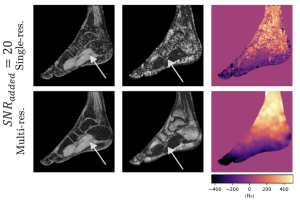
One of the main challenges in water–fat MRI is the presence of spatial variations in the main magnetic field, denoted B₀. These field inhomogeneities may arise from imperfections in the scanner’s magnetic field, imperfect shimming, or differences in magnetic susceptibility between tissues. They can become particularly large in body MRI near interfaces between air, bone, fat, and soft tissues. Their spatial distribution is described by a field map, which must be estimated together with the water and fat images.
Field inhomogeneities modify the phase of the measured MRI signal. Because this phase evolution is periodic, several field-map values may explain the same measurements almost equally well, especially when only a few echoes are acquired or when the signal-to-noise ratio (SNR) is low, as can occur in low-field MRI. Recovering the correct water image, fat image, and field map therefore requires solving a non-convex optimization problem with several competing local solutions. Selecting an incorrect solution can produce a water–fat swap, in which water is reconstructed as fat or vice versa.
Spatial regularization can reduce these errors by encouraging neighboring voxels to have consistent field-map values. Graph-cut optimization is particularly effective for selecting a spatially coherent solution. Yet, conventional approaches often rely on fixed or heuristic regularization parameters: too little regularization leaves noisy and unstable estimates, whereas too much may propagate an incorrect solution across the image.
This project will investigate a more adaptive strategy based on confidence maps. These maps will estimate how reliable the local field-map candidates are and guide the graph-cut algorithm accordingly. Reliable regions should anchor the reconstruction, while ambiguous regions should be treated more cautiously. Confidence measures may be derived from the residual landscape, the separation between competing candidates, local signal strength, and consistency across resolution levels.
The student will build on an existing in-house Python reconstruction framework containing simulation tools, voxel-wise signal fitting, candidate extraction, and multi-resolution graph-cut optimization. Simulated and real MRI datasets will be used to identify failure cases, design confidence measures, integrate them into the optimization, and assess whether they reduce water–fat swaps in challenging dual- and multi-echo acquisitions. The project therefore starts from a working codebase rather than from a blank page.
Goals:
- Reproduce and understand the existing water–fat reconstruction pipeline.
- Characterize failure modes as a function of SNR, echo timing, field strength, field map and water–fat composition.
- Design confidence maps that quantify local ambiguity in field-map estimation.
- Develop confidence-guided graph-cut regularization and compare it with the current method.
- Evaluate the reconstruction quantitatively and visually on simulated and real MRI data.
The student will have the opportunity to:
- Learn practical MRI physics, chemical-shift encoding, inverse problems, and graph-cut optimization.
- Work on an active research problem.
- Analyze real MRI data and participate in some MRI scanning session at the CHUV.
- Join the MRISM lab and interact with researchers working on MRI acquisition and reconstruction.
Requirements:
- An interest in computational imaging, signal processing, applied physics, or medical imaging.
- Good programming skills in Python; experience with Julia is an advantage but not required.
- Basic knowledge of linear algebra and numerical methods; prior MRI knowledge is helpful but not required.
- Curiosity for solving physics-driven optimization problems.
- Good command of English. French-speaking supervision is also available.
Supervisors: Louis Peyratoux, Prof. Dimitrios Karampinos
Contact details for more information: [email protected]
Relevant references:
- Alpman, A., Wei, H., and Liu, C. (2026). Reducing Fat-Water Swaps and Field Map Artifacts via Echo Dependent Phase Reconstruction for Liver QSM. In Cape Town – 2026 ISMRM-ISMRT Annual Meeting and Exhibition, Cape Town, South Africa. Program Number: 466-02-014. URL: http://echo.ismrm.org/abstracts/view/4aa73f3f-3a74-4488-8f39-1132ed8064f6.
- Eggers, H., and Boernert, P. (2014). Chemical shift encoding-based water-fat separation methods. Journal of Magnetic Resonance Imaging, 40(2), 251-268. doi: 10.1002/jmri.24568.
- Ruschke, S., Zoellner, C., Boehm, C., Diefenbach, M. N., and Karampinos, D. C. (2022). Chapter 14 – Chemical Shift Encoding-Based Water-Fat Separation. In M. Akcakaya, M. Doneva, and C. Prieto (Eds.), Magnetic Resonance Image Reconstruction, Advances in Magnetic Resonance Technology and Applications, Vol. 7, pp. 391-418. Academic Press. doi: 10.1016/B978-0-12-822726-8.00025-7.
- Bydder, M., Yokoo, T., Yu, H., Carl, M., Reeder, S. B., and Sirlin, C. B. (2011). Constraining the initial phase in water-fat separation. Magnetic Resonance Imaging, 29(2), 216-221. doi: 10.1016/j.mri.2010.08.011.
Magnetic Resonance Imaging at 3T is widely used for both anatomical and functional brain imaging, and it is increasingly important in the development and validation of novel neurotechnology. At the same time, the presence of implanted neurodevices introduces complex electromagnetic interactions with the MRI transmit field, which can lead to local field distortions, RF heating, and specific absorption rate (SAR) hotspots. Ensuring implant safety in these environments is therefore a critical step in the translation of new neuroimplants into clinical use.
In collaboration with the Laboratory for Soft Bioelectronic Interfaces (LSBI) at EPFL, which specializes in neuroengineering and implantable systems, we are establishing a comprehensive in silico simulation framework to evaluate implant safety and performance inside a 3T MRI environment. This project focuses on high-fidelity electromagnetic modeling of implants within a realistic MRI setup, including body coil excitation and human voxel models.
Goals:
- Develop and validate full-wave electromagnetic simulation models of neuroimplants inside a 3T MRI environment, including realistic body coil excitation and human voxel/phantom models.
- Perform large-scale parameter sweeps using Sim4Life to evaluate RF-induced E-fields, local SAR distributions, and identify potential safety-critical hotspots under different implant configurations and positions.
What you will learn:
- Hands-on experience with computational electromagnetics using state-of-the-art simulation software (Sim4Life).
- Practical understanding of RF safety in MRI, including SAR, E-field interactions, and implant-induced field perturbations.
- Experience working with anatomical voxel models and MRI RF coil models, including setup, meshing, and boundary conditions.
- Skills in large-scale simulation studies, parameter optimization, and data analysis in a biomedical engineering context.
Requirements:
- Strong motivation to work in computational physics, biomedical engineering, electromagnetics, or medical imaging research.
- Background in physics, electrical engineering, biomedical engineering, or a closely related field.
- Curiosity, reliability, and ability to work independently while also collaborating effectively within a small research team.
- Good command of English
Supervisors: Dr. Daniel Wenz (CIBM), Dr. Victor Druet (LSBI), Prof. Dimitrios Karampinos (MRISM)
Contact details for more information: [email protected]