At the intersection of life sciences, imaging and art, VOIR EST SAVOIR celebrates the richness of scientific imaging and the beauty of sciences over many scales

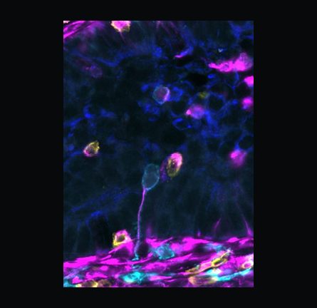
Moonwalking boutons
Synaptic boutons of the Moonwalker Descending Neurons in the fly Ventral Nerve Cord (VNC, the equivalent of the spinal cord). When these 4 neurons, which send commands from the brain to the VNC, are activated, the release of neurotransmitters from these boutons triggers a cascade of local neuron activity that drives the fly to walk backwards – or do the moonwalk!
Maite Azcorra Sedano, Postdoc, Ramdya Lab.
I investigate how the simple input from the Moonwalker Neurons is transformed by a handful of neurons into a complex, coordinated behavior.

Pre-historic get-together
The amoebo-flagellate Naegleria gruberi can rapidly and synchronously differentiate from an amoeba to a fleagellate. Here, Naegleria is celebrating and showcasing its ability to do precisely that: building de novo centrioles, flagella, and a microtubule cytoskeleton in 90 minutes, with perfection!
Dessislava Ilieva, PhD student, Gönczy Lab.
Dessislava Ilieva, PhD student at the Gönczy Lab. I am working on the unicellular organism Naegleria gruberi and more specifically on de novo centriole formation.

Whispers from the Molecular Abyss
In the silent depths of the molecular world, the spectral shell of apoferritin emerges, revealed in shadowed detail by cryo-electron microscopy. A cathedral of protein, cloaked in frost and darkness, its symmetry echoes life’s iron keepers. This journey beyond the visible was made possible with the Dubochet Center of Imaging, where technology and vision converge to uncover the finest details of life, once sealed in silence.
Yoan Duhoo, Scientist, Protein Production and Structure Core Facility.
I’m an expert in cryoEM and biophysics. Passionate about unseen science, protein purification, and supporting researchers with advanced structural biology expertise.
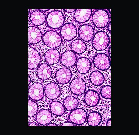
Blossoms of the Bowel
“Blossoms of the Bowel” presents an H&E-stained longitudinal section of healthy human intestine. Part of a study on colorectal cancer, this image captures the natural beauty and organized structure of healthy tissue; features that are gradually lost as cancer develops and the architecture becomes increasingly chaotic.
Elia Escoffier, PhD student, Radtke Lab.
I’m studying colorectal cancer to better understand how tumors evolve and to help design more effective therapies.

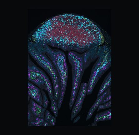
Mini Trachea, Maximum Hope
3D reconstruction of a rabbit trachea organoid derived from stem cells, imaged by confocal microscopy after tissue clearing. Nuclei (blue), ciliated cells (white), and mucus-producing goblet cells (red) reveal a functional airway structure. This preclinical model supports the development of new tracheal repair strategies.
Laure Garnier, Organoid Culture & 3D Models Expert, Bioengineering & Organoids.
I work at the Bioengineering and Organoids Platform, where we support research teams in developing advanced 3D models to drive innovation in both fundamental biomedical and translational research.

Molecular Portraits
A 3D rendering of the molecular landscape of human spinal cord injury, captured through spatial transcriptomics. Each cylinder marks a spatial barcode, scaled by cell type signature. The top section represents the lesion epicenter; the bottom, intact tissue below the injury. From top left to bottom right, the shifting landscapes of neurons, immune cells, oligodendrocytes, and astrocytes reveal the dramatic reorganization of cellular composition after trauma.
Matthieu Gautier, Postdoc, .Neurorestore.
I study the molecular logic driving the cellular response to spinal cord injury to uncover new strategies for repair.

What pulls the strings?
In a single drop of lake water, entire worlds unfold, full of single-celled species that hunt, flee, and interact with seeming purpose. Among them is Didinium nasutum, a predator whose agility rivals that of macroscopic hunters. This image reveals the acetylated tubulin scaffolds (magenta) on its body and the hair-like cilia (green) it uses to swim. Yet we see no brain. How do these filaments coordinate motion so precisely and what pulls the strings?
Katerina Kourkoulou, PhD student, Living Patterns Lab.
We investigate how biological patterns arise from microscopic interactions. I explore how coordinated ciliary beating fine-tunes flow allowing emergent behavior.

There is fire in your eyes
The image illustrates the expression of the immune effector molecule DiptericinB, in the degenerating eyes (note the black eye lesions) of the fruit fly, Drosophila melanogaster.
Kenan Krakovic, PhD student, Lemaitre Lab.
I work on neurodegeneration and immunity using fruit flies as a model.
Gold Medal

Holed Hela
HeLa cells were incubated in 384-well plates with bacteria (Chlamydia trachomatis), forming large inclusions in infected cells. 5600 molecules were screened to identify those that prevent infection. The goal is deciphering Rab-based endocytosis (collaboration with E Trofimenko, UNIL). Nuclei are in cyan. An in-house far-red dye highlights the cytoplasm (magenta), emphasizing bacterial inclusions, which appear as unstained voids deforming the cell. Can you spot which cells escaped infection?
Fabien Kuttler, Scientist, Biomolecular Screening Core Facility.
I am a scientist in High Content Screening and manage image-based screening projects using automated fluorescent microscopy for the EPFL PTCB.

Wired to swim
This image shows the surface of a single-celled organism called Paramecium, which uses tiny hair-like cilia to swim and feed. Cilia are anchored by basal bodies (cyan), arranged in rows and connected by red fibers. The spiral-shaped structure is a contractile vacuole that pumps out excess water. The image was taken with a 40x confocal microscope and expanded for super-resolution. The area shown is just 16 × 11 microns in real size.
Daphne Laan, PhD student, Living Patterns Lab.
In our lab, we study how cilia on cells are connected and coordinated and how this translates to complex single cell behaviors.

In a galaxy not so far, far away…
Resembling a distant cosmic nebula, this image captures the intricate architecture of a miniature biological structure. Here I present a murine small intestinal organoid, a three-dimensional tissue model derived from stem cells that mimics key features of the native intestine. In this image, the apical surface of the organoid is shown in blue, while subcellular organelles are marked by bright spots within the tissue.
Jakob J. Langer, Postdoctoral researcher, Lutolf Lab.
I am a researcher with a particular interest in developing sophisticated miniature-organs capable of capturing the biological intricacies of pathophysiological processes.
Gold Medal

Surfing waves of the unseen world
Mutant Pseudomonas aeruginosa bacteria form collective waves, revealed by AI-tracked trajectories in this phase contrast image. Lacking a surface-sensing gene, they fail to reverse upon collisions with their neighbours — unlike their wild-type peers — shifting from diffusive to directed motion. It reminds us that even a small change in individual behavior can reshape the dynamics of the community — bacterial or human alike.
Laure Le Blanc, Postdoc, Persat Lab.
I’m passionate about bacterial communities and microbiomes and I use interdisciplinary approaches to reveal bacteria’s remarkable adaptability across time and space.

Gastruloids in full bloom
Gastruloids are 3D in vitro models of early development. From homogeneous cell populations, they can self-organize and autonomously differentiate into diverse cell types, forming complex structures and internal cavities. In this image, fluorescent markers reveal one of these emerging cell identities, offering a glimpse into the dynamic processes shaping early life.
Maxine Leonardi, PhD student, Naef Lab.
I am studying the interplay between cell cycle dynamics and embryonic development using mouse gastruloids as a model.
Bronze medal

Fire explosion
Immunofluorescence confocal imaging of a Paramecium cilia and trychocyst exocytosis. When we change the calcium concentration of the solution, Paramecia uses specialized organelles called trichocysts as a rapid defense mechanism. These elongated, capsule-like structures are embedded in the outer layer of the cell and can be explosively discharged, forming a spiky barrier around the organism. The immunofixation process can also alter the ciliation along the surface, usually fully covered.
Benedetta Noferi, PhD student, Living Patterns Lab.
I aim to study the complex world of unicellular eukaryotes and their cellular biophysical mechanisms!

Sanctuary for Survival
This 3D reconstruction of an SBFSEM image shows uropathogenic E. coli (yellow) persisting in urothelial niches (purple, nuclei; transparent, cell membrane) as cell wall deficient forms. In our bladder-on-chip model, antibiotic treatment in high-osmolarity urine triggered this form switching from the classic bacterial rod shape. This allowed UPEC to persist within the tissue and cause recurrent infections.
Gauri Paduthol, PhD student, McKinney Lab.
I am developing a bladder organ-on-chip model to investigate recurrent urinary tract infections. This image was constructed together with the BioEM team.
Kissing balloons
Two immune cells, a migrating cyan macrophage, and a yellow CD4 T cell, meet in a colorectal mouse tissue. Images like these allow the study of tissue organization and cell interaction. The macrophage’s tail is purple with vimentin, a protein providing support and flexibility in its movement. These two cell types are known to interact, either to activate the T cell or to induce its death. Here their close contact raises the question: is this an activating kiss of life, or a fatal kiss of death?
Anjalie Schlaeppi, System Specialist, Histology and Biooptics Core Facilities.
After a PhD in light sheet microscopy, I implement spatial multi-omics at the Histology and BIOP platforms. I’m interested in finding interesting details in tissue architecture, and teaching.

Opening windows into the cell
This image shows a cellular lamella—a thin slice of a cell prepared using focused ion beam milling. This technique preserves the native architecture of the cell while making it thin enough for cryo-electron tomography. The sample is vitrified, avoiding ice crystal formation thanks to a method pioneered by Nobel laureate Jacques Dubochet. If you look closely, you’ll notice a large hexagonal ice crystal in the background.
Jonathan Schneider & Anna-Sophia Krebs, Cryo-EM scientists at the Thomä lab and the Protein Production and Structure Core Facility.
Made in collaboration with the Electron Microscopy Facility at UNIL and the Dubochet Center for Imaging.
Medusa
A medley of immune cells in a mouse small intestine evokes a Medusa-like form. At the top, red B cells cluster in a Peyer’s patch, like a jellyfish head, surrounded by cyan CD4 T cells. Patches like these are immune system centers of the intestine. Below, magenta CD8 T cells and green macrophages patrol the villi, like trailing arms, ready to respond to threats. A blue and yellow backdrop reveals overall tissue structure. Images like these allow the study tissue organization.
Jessica Sordet, Head of the Histology Core Facility.
I am a biologist passionate about histology, immunofluorescence, and omics, leading EPFL’s Histology Unit since 2007.

Gotta grab the nucleus!
The protein centrin is commonly known for its localization to the centrioles of the cell. However, in early lineage eukaryotes, centrin also forms diverse structural assemblies. In my project, I aim to understand the role of the various structures formed by centrin in the green algae Chlamydomonas. Here, I look at Chlamydomonas cells using Expansion microscopy to identify the unique structures formed by centrin (magenta) which connect the base of the cilia (cyan) to the nucleus (yellow).
Aparna Sudhakar, Postdoc, Living Patterns Laboratory.
I am interested in understanding structural assemblies formed by centrin in unicellular eukaryotes.

Order conceals disorder
This micropatterned array allows to grow intestinal organoids in a standardized manner. We make use of such a system to look at the circadian rhythm of each organoid and at their development with immunofluorescence. When these mini-organs do not have a regular circadian clock, however, they look all different: therefore they look all aligned and ordered but inside they show disordered patterns.
Elena Tonin and Florian Curvaia, PhD students, Naef Lab.
I work on the interaction between circadian clock and cell cycle in intestinal organoids, via micropatterned arrays.

Label-free lipids on a chip!
Quantitative phase images (QPI) of adipose stem and progenitor cells (ASPCs) in a microfluidic channel as they are forming lipid droplets (yellow and red) – differentiating into adipocytes. Lipids have a much higher refractive index than the cytosol of cells (nlipids~1.47 > ncyto~1.37). This creates a strong phase shift in the illumination field that an interference hologram can capture!
Jose Vasquez Porto Viso, PhD student, Deplancke Lab.
I am working on developing technologies for label-free, single-cell phenotyping!