Scientific Assistant: Farzad Rezaeianaran
Office: BM3130, E-mail: [email protected]
Project Type: Preferably Master Thesis or Semester Project
Project Section: Microengineering
Time frame: Master Thesis (4-6 months full-time), Semester Project (14 weeks, ~7h/week),
Official Start Date: 2022
Description: The laboratory of microsystems 2 is offering a master (thesis or semester) project on high-resolution in vivo imaging of bacteria in the Caenorhabditis elegans (C. elegans) gut. C. elegans is a versatile genetically tractable model organism for studying gut bacterial colonization and its influence on the health and ageing of the host [1]. High-resolution imaging of this organism requires good immobilization, prime examples of which can be seen in microfluidic devices reported in [2] (valve-based immobilization) and [3] (using trapping channels and valves). However, these devices may be less suited for long-term studies on gut bacterial colonization and indeed there are no publications on high-resolution imaging of the gut microbiota. Therefore, the aim of this master (thesis or semester) project is to fill this gap present in microfluidic technologies as well as microbiota research by enabling on-chip imaging of the worm microbiota at single-bacterium resolution.
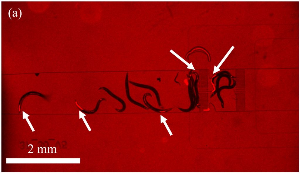
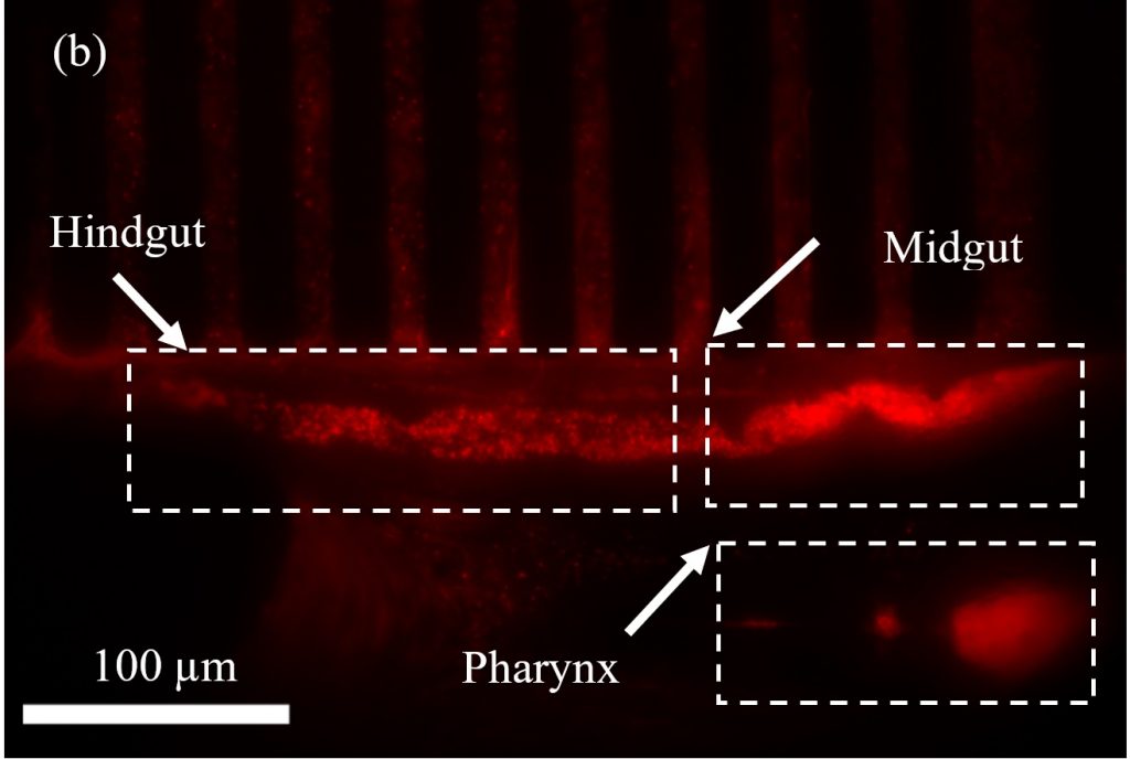
Figure 1. Widefield fluorescence microscopy graph (in the Red Fluorescence Protein (RFP) channel) of 3rd day adult wild-type C. elegans nematodes, fed with RFP E. coli OP50. (a) Freely moving worms in the microfluidic chip (b) Resolved bacteria can be seen in the hindgut of the worm immobilized in the chip.
Student Gain: The student (master thesis or semester project*) will gain a multidisciplinary experience in clean room microfabrication, microfluidics, simulation, optical imaging and image analysis.
- Clean room activities involving mask fabrication, SU-8 processing, PDMS chip fabrication
- Physics simulation of flow profiles for the microfluidic device using COMSOL
- Testing the performance of the fabricated chips for the immobilization of C. elegans involving operating the microfluidic chip, optical imaging and image analysis.
Required skills:
- Familiarity with microfabrication processes and microfluidics
- Experience in EPFL cleanroom is a strong plus
- Preferably, basic knowledge of Comsol simulation
- Basic lab skills such as solution preparation, dilutions, pipetting, etc.
For those interested in master thesis/semester project or need more information, please contact us at
[email protected]
*it should be noted that due to time constraints, the scope of a semester project will be limited to a selection of the mentioned tasks.
[1] Cabreiro, F., & Gems, D. (2013). EMBO molecular medicine, 5(9), 1300-1310. https://doi.org/10.1002/emmm.201100972
[2] Keil, W., et al. (2017). Developmental cell, 40(2), 202-214. https://doi.org/10.1016/j.devcel.2016.11.022
[3] Berger, et al. (2018). Lab on a Chip, 18(9), 1359-1368. https://doi.org/10.1039/C7LC01185G
Scientific Assistant: Muaz Draz e-mail: [email protected]
Project Type: Master Thesis/Semester Project Section: Microengineering
Timeframe: Master Thesis (5-6 months full-time), Semester Project (2-4 months part-time),
Official Start Date: 2021/2022
Description: The Laboratory of Microsystems 2 is offering a master (semester) project related to the development of microfluidic systems for cancer diagnostics. The lab has long history with developing microfluidic systems for Immunohistochemistry (IHC) and In Situ Hybridization (ISH) techniques for rapid, accurate, and costeffective cancer diagnostics. Several references [1, 2] give an overview of the work done. Simply, a tissue biological sample is loaded on a charged glass slide which is then incubated inside a microfluidic system that handles all the fluidic delivery and waste. See Figure 1 (left & right). Immunoreactions such as IHC and ISH inside microfluidic systems usually suffer from diffusion-limited transport of bioreagents inside the reaction chamber. Recently, we have been working on integrating AC elektrokinetic mixing technologies [3] that enhance the accuracy and improve the speed while possibly reducing the cost of such analysis.
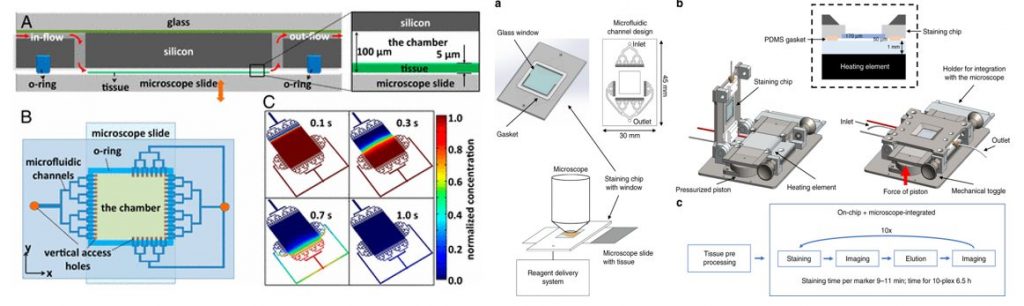
Figure 1. Left: microfluidic system & chamber design with fluid handling [1] Right: The whole microfluidic tissue processor device [2].
Student Gain: The student (master or semester project) will gain a multidisciplinary experience in microfluidic chip realization, simulation, image analysis, cancer diagnostic techniques and bio-image analysis.
- Simulation: Physics simulation of AC elektrokinetics and correlation with experimental results.
- Integrating glass substrates into the microfluidic tissue processor and setting up electronic connections.
- Carrying out IHC and ISH experiments on biological samples and results analysis.
- For a master project, possibility to carry out clean room fabrication.
Required skills:
- Understanding of clean room fabrication methods and microfluidics basics.
- Preferably, basic knowledge of Comsol simulation.
- Basic lab skills such as buffer preparation, dilutions, pipetting…. etc
For those interested for master/semester project or need more information, please contact us at [email protected]
[1] Tuna et al, PNAS April 2, 2013 110 (14) 5363-5368, https://doi.org/10.1073/pnas.1211273110
[2] Migliozzi, D. et al. Microsyst Nanoeng 5, 59 (2019). https://doi.org/10.1038/s41378-019-0104-z.
[3] H. C. Feldman , M. Sigurdson and C. D. Meinhart , Lab Chip, 2007, 7 , 1553 —1559, https://doi.org/10.1039/B706745C
Timeframe
Master Thesis (5-6 months full-time), Internship (2-6 months full-time) or Semester Project (2-4 months part-time)
Contact
Fabien Tâche ([email protected])
Description
The Laboratory of Microsystems (LMIS2) is developing an innovative laboratory device for drug discovery research. The technology employs robotics and computer vision for the automated culture and analysis of micro-organisms within microfluidic chips, as powerful biological models to investigate human diseases and test their potential cures. In this framework, we look for students to contribute to the development of the following microfluidics components:
• Highly-integrated distribution valves with a high number of outputs
• High resolution syringe pumps
• New generation of microfluidic chips, including the optimization of the manufacturing process
The aim of this project is to design one (or several) of the devices listed above, manufacture them, and characterize their performances. If the implementation is successful, the devices will be integrated in the existing robotic platform and used for experiments on C. elegans.
The overall project holds great potential to represent a breakthrough in biomedical research and, as such, resides at the core of a startup project (Nagi Bioscience).
Timeframe
Master Thesis (5-6 months full-time), Internship (2-6 months full-time) or Semester Project (2-4 months part-time)
Contact
Fabien Tâche ([email protected])
Description
The Laboratory of Microsystems (LMIS2) is developing an innovative laboratory device for drug discovery research. The technology employs robotics and computer vision for the automated culture and analysis of micro-organisms within microfluidic chips, as powerful biological models to investigate human diseases and test their potential cures. In this framework, we look for students to contribute to the development of experimental protocols and run biological assays. This includes the following tasks:
• Definition of experimental protocols
• Implementation and optimization of protocols (manipulation of C. elegans using microfluidics, feeding of worms, control of worm chamber temperature, …)
• Perform biological assays to validate the technology and protocols
This project is highly multidisciplinary, because it is at the interface of technology, life sciences and entrepreneurship.
The overall project holds great potential to represent a breakthrough in biomedical research and, as such, resides at the core of a startup project (Nagi Bioscience).
Timeframe
Master Thesis (5-6 months full-time), Internship (2-6 months full-time) or Semester Project (2-4 months part-time)
Contact
Fabien Tâche ([email protected])
Description
The Laboratory of Microsystems (LMIS2) is developing an innovative laboratory device for drug discovery research. The technology employs robotics and computer vision for the automated culture and analysis of micro-organisms within microfluidic chips, as powerful biological models to investigate human diseases and test their potential cures. In this framework, we look for students to contribute to the development of a tracking and analysis software. This project includes the following tasks:
• Implementation of worm tracking algorithms
• Implementation of a worm phenotype analysis software
• Characterization, optimization and validation of algorithms
• Implementation of a graphical user interface (GUI) displaying the results of automated analysis
This project requires good software development and image processing skills. Machine learning knowledge is a plus.
The overall project holds great potential to represent a breakthrough in biomedical research and, as such, resides at the core of a startup project (Nagi Bioscience).